Glucosidase/Rutinosidase from Mucor circinelloides, Penicillium chrysogenum and Aspergillus niger

Jan Stransky.

SASDVT8 – Glucosidase/Rutinosidase from Aspergillus niger - glycosylated

glucan 1,3-beta-glucosidase (W90L, N179H)
MWI(0) 85 kDa
MWexpected 83 kDa
VPorod 111 nm3
log I(s) 6.10×10⁻² 6.10×10⁻³ 6.10×10⁻⁴ 6.10×10⁻⁵
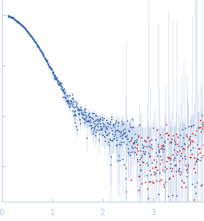
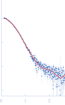
glucan 1,3-beta-glucosidase (W90L, N179H) small angle scattering data  s, nm-1
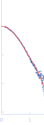
ln I(s)
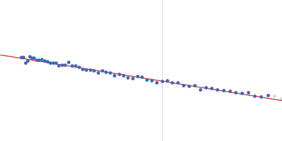
glucan 1,3-beta-glucosidase (W90L, N179H) Guinier plot ln 6.11×10⁻² Rg: 3.0 nm 0 (3.0 nm)-2 s2
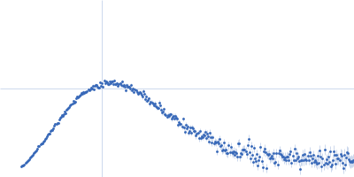
(sRg)2I(s)/I(0)
glucan 1,3-beta-glucosidase (W90L, N179H) Kratky plot 1.104 0 √3 sRg
p(r)
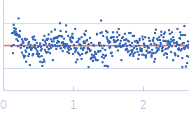
glucan 1,3-beta-glucosidase (W90L, N179H) pair distance distribution function Rg: 2.9 nm 0 Dmax: 8.1 nm

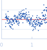
Data validation


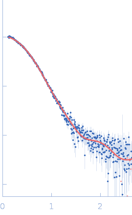
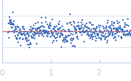
Fits and models


log I(s)
 s, nm-1
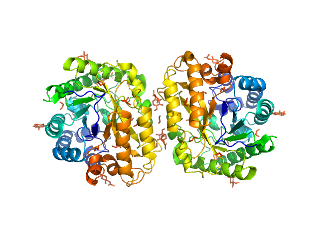
glucan 1,3-beta-glucosidase (W90L, N179H) PDB (PROTEIN DATA BANK) model

log I(s)
 s, nm-1
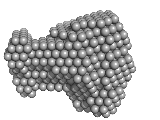
glucan 1,3-beta-glucosidase (W90L, N179H) DAMMIN model

log I(s)
 s, nm-1

SAXS data from solutions of Glucosidase/Rutinosidase (glycosylated) in McIlvain (Na2HPO4/citric acid) buffer, pH 3.5 were collected using an Anton Paar SAXSpoint 2.0 instrument at the Institute of Biotechnology, Czech Academy of Sciences/Centre of Molecular Structure (Vestec, Czech Republic) equipped with an Eiger R 1M detector at a sample-detector distance of 0.8 m and at a wavelength of λ = 0.134 nm (I(s) vs s, where s = 4πsinθ/λ, and 2θ is the scattering angle). One solute concentration of 3.40 mg/ml was measured at 20°C. 40 successive 30 second frames were collected. The data were normalized to the intensity of the transmitted beam and radially averaged; the scattering of the solvent-blank was subtracted.

Raw data are available at: https://doi.org/10.5281/zenodo.13950823

glucan 1,3-beta-glucosidase (W90L, N179H) (AnRut)
Mol. type   Protein
Organism   Aspergillus niger (strain ATCC 1015 / CBS 113.46 / FGSC A1144 / LSHB Ac4 / NCTC 3858a / NRRL 328 / USDA 3528.7)
Olig. state   Dimer
Mon. MW   41.6 kDa
 
UniProt   G3YFQ1 (20-386)
Sequence   FASTA
 
PDB ID   6I1A