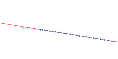
Molecular structure and enzymatic mechanism of the human collagen hydroxylysine galactosyltransferase GLT25D1/COLGALT1

De Marco M, Rai S, Scietti L, Mattoteia D, Liberi S, Moroni E, Pinnola A, Vetrano A, Iacobucci C, Santambrogio C, Colombo G, Forneris F, Nature Communications 16(1) (2025) DOI

SASDVZ2 – Human Collagen Galactosyltransferase GLT25D1/COLGALT1

Procollagen galactosyltransferase 1
MWI(0) 131 kDa
MWexpected 137 kDa
VPorod 184 nm3
log I(s) 2.29×101 2.29×100 2.29×10⁻¹ 2.29×10⁻²
Procollagen galactosyltransferase 1 small angle scattering data  s, nm-1
ln I(s)
Procollagen galactosyltransferase 1 Guinier plot ln 2.29×101 Rg: 4.3 nm 0 (4.3 nm)-2 s2
(sRg)2I(s)/I(0)
Procollagen galactosyltransferase 1 Kratky plot 1.104 0 3 sRg
p(r)
Procollagen galactosyltransferase 1 pair distance distribution function 0 Dmax: 14.0 nm

Data validation


Fits and models


log I(s)
 s, nm-1
Procollagen galactosyltransferase 1 CORAL model

Synchrotron SAXS data from solutions of Human Collagen Galactosyltransferase GLT25D1/COLGALT1 in 25 mM HEPES, 0.1 M NaCl, pH 8 were collected on the BM29 beam line at the ESRF storage ring (Grenoble, France) using a Pilatus3 2M detector at a sample-detector distance of 2.9 m and at a wavelength of λ = 0.099187 nm (I(s) vs s, where s = 4πsinθ/λ, and 2θ is the scattering angle). In-line size-exclusion chromatography (SEC) SAS was employed. The SEC parameters were as follows: A 50.00 μl sample at 4 mg/ml was injected at a 0.06 ml/min flow rate onto a GE Superdex 200 Increase 3.2/300 column at 20°C. 1200 successive 1 second frames were collected. The data were normalized to the intensity of the transmitted beam and radially averaged; the scattering of the solvent-blank was subtracted.
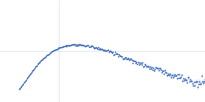
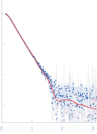
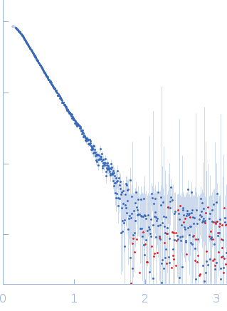
Procollagen galactosyltransferase 1 (GLT25D1)
Mol. type   Protein
Organism   Homo sapiens
Olig. state   Dimer
Mon. MW   68.5 kDa
 
UniProt   Q8NBJ5 (30-618)
Sequence   FASTA