|
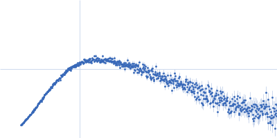
Synchrotron SAXS
data from solutions of
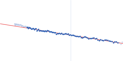
DGSt - Chimeric protein consisting of DCV, GFP and SpyTag domain
in
20 mM Tris, 150 mM NaCL, 3% glycerol, pH 7.5
were collected
on the
EMBL P12 beam line
at the PETRA III storage ring
(DESY; Hamburg, Germany)
using a Pilatus 6M detector
at a sample-detector distance of 3 m and
at a wavelength of λ = 0.123985 nm
(I(s) vs s, where s = 4πsinθ/λ, and 2θ is the scattering angle).
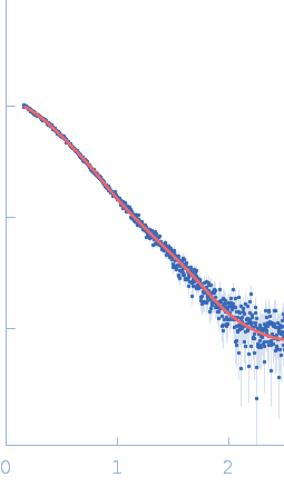
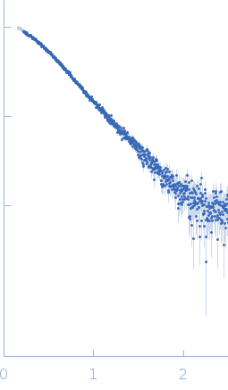
In-line size-exclusion chromatography (SEC) SAS was employed. The SEC parameters were as follows: A 80.00 μl sample
at 8 mg/ml was injected at a 0.70 ml/min flow rate
onto a Cytiva Superdex 200 Increase 10/300 column
at 20°C.
3000 successive
1 second frames were collected.
The data were normalized to the intensity of the transmitted beam and radially averaged; the scattering of the solvent-blank was subtracted.
Note: The experimentally determined protein mass (SEC-MALS) is overestimated due to the presence of a fluorescent protein in the sample. The light scattering laser operates at a wavelength that excites the fluorescent protein, leading to inaccurate signal measurements.
|
|
DCV-GFP-St
(DGSt)
|
| Mol. type |
|
Protein |
| Organism |
|
synthetic construct |
| Olig. state |
|
Monomer |
| Mon. MW |
|
41.3 kDa |
| Sequence |
|
FASTA |
| |
|
 s, nm-1