A ternary switch model governing ERα ligand binding domain conformation.

McDougal DP, Pederick JL, Novick SJ, Jovcevski B, Warrender AK, Pascal BD, Griffin PR, Bruning JB, Nat Commun 16(1):10363 (2025) Europe PMC

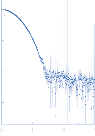
SASDWD7 – SEC-SAXS data of wildtype Melanotaenia fluviatilis apo estrogen receptor alpha ligand binding domain (LBD) at pH 7.5

Estrogen receptor alpha
MWexperimental 58 kDa
MWexpected 57 kDa
VPorod 72 nm3
log I(s) 8.29×10⁻³ 8.29×10⁻⁴ 8.29×10⁻⁵ 8.29×10⁻⁶
Estrogen receptor alpha small angle scattering data  s, nm-1
ln I(s)
Estrogen receptor alpha Guinier plot ln 8.30×10⁻³ Rg: 2.5 nm 0 (2.5 nm)-2 s2
(sRg)2I(s)/I(0)
Estrogen receptor alpha Kratky plot 1.104 0 3 sRg
p(r)
Estrogen receptor alpha pair distance distribution function Rg: 2.5 nm 0 Dmax: 9.2 nm

Data validation


Fits and models


log I(s)
 s, nm-1
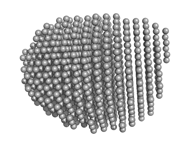
Estrogen receptor alpha DAMMIF model

Synchrotron SAXS data from solutions of SEC-SAXS data of wildtype Melanotaenia fluviatilis apo estrogen receptor alpha ligand binding domain (LBD) at pH 7.5 in 20 mM Tris-HCl pH 7.5, 150 mM NaCl, 5% glycerol, 2 mM TCEP, pH 7.5 were collected on the BioSAXS beam line at the Australian Synchrotron storage ring (Melbourne, Australia) using a Pilatus3 X 2M detector at a sample-detector distance of 2.2 m and at a wavelength of λ = 0.1 nm (I(s) vs s, where s = 4πsinθ/λ, and 2θ is the scattering angle). In-line size-exclusion chromatography (SEC) SAS was employed. The SEC parameters were as follows: A 80.00 μl sample at 5 mg/ml was injected at a 0.30 ml/min flow rate onto a column at 19.8°C. 641 successive 1 second frames were collected. The data were normalized to the intensity of the transmitted beam and radially averaged; the scattering of the solvent-blank was subtracted.

SEC column = UNKNOWN

Estrogen receptor alpha (ERalpha)
Mol. type   Protein
Organism   Melanotaenia fluviatilis
Olig. state   Dimer
Mon. MW   28.7 kDa
 
UniProt   D6N7U3 (310-553)
Sequence   FASTA