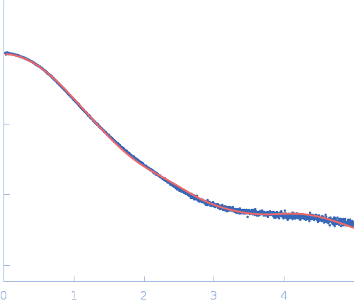
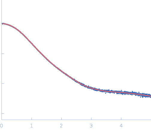
Crystal structure of the class V GTP-binding RNA aptamer bound to its ligand: GTP recognition by a topologically complex intermolecular G-quadruplex.

Stafflinger H, Neißner K, Bartsch S, Pichler AK, Bartosik K, Dhamotharan K, Abele R, Duchardt-Ferner E, Micura R, Schindelin H, Wöhnert J, Nucleic Acids Res 53(22) (2025) Europe PMC

SASDWJ4 – Class V GTP aptamer complex with GTP

Class V GTP aptamer
Guanosine triphosphate
MWexperimental 24 kDa
MWexpected 23 kDa
VPorod 34 nm3
log I(s) 5.67×10⁻¹ 5.67×10⁻² 5.67×10⁻³ 5.67×10⁻⁴
Class V GTP aptamer Guanosine triphosphate small angle scattering data  s, nm-1
ln I(s)
Class V GTP aptamer Guanosine triphosphate Guinier plot ln 5.68×10⁻¹ Rg: 2.2 nm 0 (2.2 nm)-2 s2
(sRg)2I(s)/I(0)
Class V GTP aptamer Guanosine triphosphate Kratky plot 1.104 0 3 sRg
p(r)
Class V GTP aptamer Guanosine triphosphate pair distance distribution function Rg: 2.3 nm 0 Dmax: 7.6 nm

Data validation


Fits and models


log I(s)
 s, nm-1
Class V GTP aptamer Guanosine triphosphate COOT model

log I(s)
 s, nm-1
Class V GTP aptamer Guanosine triphosphate DAMMIN model

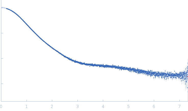
Synchrotron SAXS data from solutions of Class V GTP aptamer complex with GTP in 50 mM Bis-Tris, 50 mM KCl, 2 mM MgCl2, pH 6.5 were collected on the EMBL P12 beam line at PETRA III (DESY; Hamburg, Germany) using a Pilatus 6M detector at a sample-detector distance of 3 m and at a wavelength of λ = 0.123985 nm (I(s) vs s, where s = 4πsinθ/λ, and 2θ is the scattering angle). One solute concentration of 6.00 mg/ml was measured at 20°C. 40 successive 0.095 second frames were collected. The data were normalized to the intensity of the transmitted beam and radially averaged; the scattering of the solvent-blank was subtracted.

Class V GTP aptamer
Mol. type   RNA
Organism   Escherichia coli
Olig. state   Monomer
Mon. MW   22.1 kDa
Sequence   FASTA
 
Guanosine triphosphate (GTP)
Mol. type   Other
Olig. state   Monomer
Mon. MW   0.5 kDa
Chemical formula