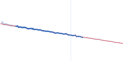
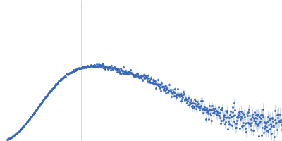
Unique conformational dynamics and protein recognition of A-to-I hyper-edited dsRNA.

Müller-Hermes C, Piomponi V, Hilber S, Asami S, Kreutz C, Bussi G, Sattler M, Nucleic Acids Res 53(12) (2025) Europe PMC

SASDWM5 – A-RNA, non-edited 20mer dsRNA

A-RNA, Top-Strand
A-RNA, Bottom-Strand
MWexperimental 13 kDa
MWexpected 13 kDa
VPorod 17 nm3
log I(s) 1.31×101 1.31×100 1.31×10⁻¹ 1.31×10⁻²
A-RNA, Top-Strand A-RNA, Bottom-Strand small angle scattering data  s, nm-1
ln I(s)
A-RNA, Top-Strand A-RNA, Bottom-Strand Guinier plot ln 1.32×101 Rg: 1.8 nm 0 (1.8 nm)-2 s2
(sRg)2I(s)/I(0)
A-RNA, Top-Strand A-RNA, Bottom-Strand Kratky plot 1.104 0 3 sRg
p(r)
A-RNA, Top-Strand A-RNA, Bottom-Strand pair distance distribution function Rg: 1.9 nm 0 Dmax: 6.2 nm

Data validation


There are no models related to this curve.

Synchrotron SAXS data from solutions of A-RNA, non-edited 20mer dsRNA in 25 mM sodium phosphate 25 mM sodium chloride, pH 6.4 were collected on the BM29 beam line at the ESRF storage ring (Grenoble, France) using a Pilatus3 2M detector at a wavelength of λ = 0.09919 nm (I(s) vs s, where s = 4πsinθ/λ, and 2θ is the scattering angle). In-line size-exclusion chromatography (SEC) SAS was employed. The SEC parameters were as follows: A 80.00 μl sample at 2 mg/ml was injected at a 0.20 ml/min flow rate onto a GE Superdex 200 Increase 5/150 column at 20°C. 500 successive 2 second frames were collected. The data were normalized to the intensity of the transmitted beam and radially averaged; the scattering of the solvent-blank was subtracted.
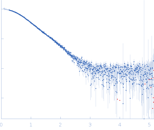
Sample detector distance = UNKNOWN

A-RNA, Top-Strand (A-RNA, TS)
Mol. type   RNA
Olig. state   Monomer
Mon. MW   6.5 kDa
Sequence   FASTA
 
A-RNA, Bottom-Strand (A-RNA, BS)
Mol. type   RNA
Olig. state   Monomer
Mon. MW   6.4 kDa
Sequence   FASTA