Conformational multiplicity of bacterial ferric binding protein revealed by small angle x-ray scattering and molecular dynamics calculations

Liu G Ekmen E, Jalalypour F, Mertens H, Jeffries C, Svergun D, Atilgan A, Atilgan C, Sayers Z The Journal of Chemical Physics 158(8) (2023) DOI

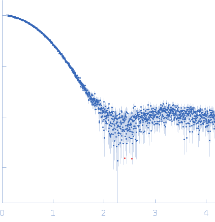
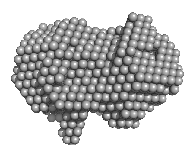
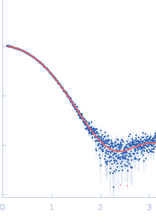
SASDWM7 – D52A mutant of ferric binding protein (FbpA) in holo (with Fe) form in low ionic strength (LIS) buffer (1/10 PBS)

Iron-utilization periplasmic protein
MWexperimental 27 kDa
MWexpected 34 kDa
VPorod 24 nm3
log I(s) 4.26×10⁻² 4.26×10⁻³ 4.26×10⁻⁴ 4.26×10⁻⁵
Iron-utilization periplasmic protein small angle scattering data  s, nm-1
ln I(s)
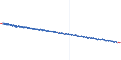
Iron-utilization periplasmic protein Guinier plot ln 4.27×10⁻² Rg: 2.0 nm 0 (2.0 nm)-2 s2
(sRg)2I(s)/I(0)
Iron-utilization periplasmic protein Kratky plot 1.104 0 3 sRg
p(r)
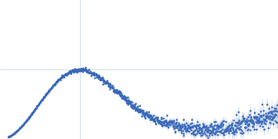
Iron-utilization periplasmic protein pair distance distribution function Rg: 2.0 nm 0 Dmax: 6.1 nm

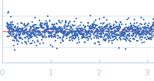
Data validation


Fits and models


log I(s)
 s, nm-1
Iron-utilization periplasmic protein DAMFILT model

Synchrotron SAXS data from solutions of D52A mutant of ferric binding protein (FbpA) in holo (with Fe) form in low ionic strength (LIS) buffer (1/10 PBS) in 1 mM Na2HPO4.7H2O, 0.18 mM KH2PO4, 13.7 mM NaCl, 0.27 mM KCl, 5%v/v Glycerol, pH 7.4 were collected on the EMBL P12 beam line at the PETRA III storage ring (DESY; Hamburg, Germany) using a Pilatus 6M detector at a sample-detector distance of 3 m and at a wavelength of λ = 0.124 nm (I(s) vs s, where s = 4πsinθ/λ, and 2θ is the scattering angle). One solute concentration of 3.00 mg/ml was measured at 4°C. 40 successive 1 second frames were collected. The data were normalized to the intensity of the transmitted beam and radially averaged; the scattering of the solvent-blank was subtracted.

Iron-utilization periplasmic protein (FBPA_HAEIN)
Mol. type   Protein
Organism   Haemophilus influenzae
Olig. state   Monomer
Mon. MW   33.8 kDa
 
UniProt   P35755 (23-332)
Sequence   FASTA