A structural basis for chaperone repression of stress signaling from the endoplasmic reticulum.

Neidhardt L, Tung J, Kuchersky M, Milczarek J, Kargas V, Stott K, Rosenzweig R, Ron D, Yan Y Mol Cell (2025) Europe PMC

SASDWU6 – Human anterior gradient protein 2 (AGR2)

Anterior gradient protein 2 homolog
MWexperimental 24 kDa
MWexpected 30 kDa
VPorod 30 nm3
log I(s) 1.77×10⁻¹ 1.77×10⁻² 1.77×10⁻³ 1.77×10⁻⁴
Anterior gradient protein 2 homolog small angle scattering data  s, nm-1
ln I(s)
Anterior gradient protein 2 homolog Guinier plot ln 1.78×10⁻¹ Rg: 2.0 nm 0 (2.0 nm)-2 s2
(sRg)2I(s)/I(0)
Anterior gradient protein 2 homolog Kratky plot 1.104 0 3 sRg
p(r)
Anterior gradient protein 2 homolog pair distance distribution function Rg: 2.0 nm 0 Dmax: 6.3 nm

Data validation


Fits and models


log I(s)
 s, nm-1
Anterior gradient protein 2 homolog MULTIFOXS model

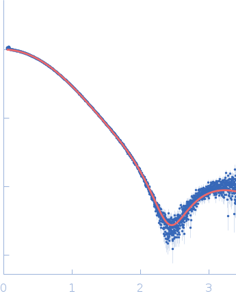
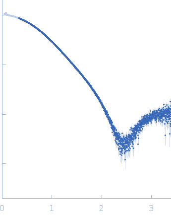
Synchrotron SAXS data from solutions of anterior gradient protein 2 (AGR2) in 20 mM Tris pH7.4, 150 mM NaCl, 1% glycerol, 0.2 mM TCEP, were collected on the B21 beam line at the Diamond Light Source (Didcot, UK) using a Eiger 4M detector at a sample-detector distance of 3.7 m and at a wavelength of λ = 0.09464 nm (I(s) vs s, where s = 4πsinθ/λ, and 2θ is the scattering angle). In-line size-exclusion chromatography (SEC) SAS was employed. The SEC parameters were as follows: A 35.00 μl sample at 46 mg/ml was injected at a 0.06 ml/min flow rate onto a GE Superdex 75 Increase 3.2/300 column at 15°C. 30 successive 1 second frames were collected through the sample elution peak (from a total of 600 frames collected throughout the entire SEC run). The data were normalized to the intensity of the transmitted beam and radially averaged; the scattering of the solvent-blank was subtracted.

Anterior gradient protein 2 homolog (AGR2)
Mol. type   Protein
Organism   Homo sapiens
Olig. state   Dimer
Mon. MW   15.1 kDa
 
UniProt   O95994 (41-171)
Sequence   FASTA