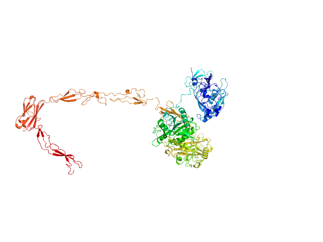
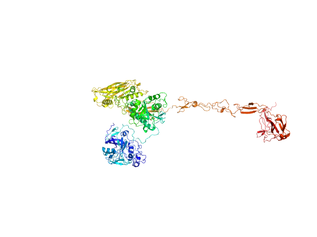
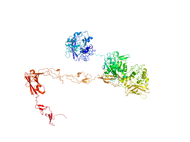
Thomasclavelia ramosa IgA peptidase (IgAse) active site mutant (A31-I1166)

Juan Sebastián Ramírez Larrota.

SASDX42 – IgA peptidase (A31-I1166; active site mutant E540A)

IgA protease (E540A)
MWexperimental 119 kDa
MWexpected 128 kDa
VPorod 190 nm3
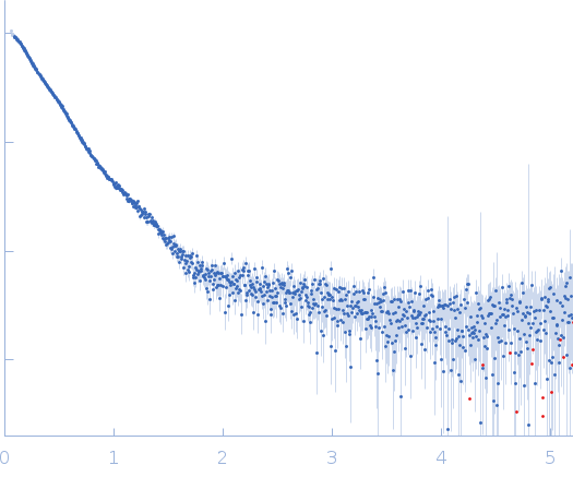
log I(s) 7.01×101 7.01×100 7.01×10⁻¹ 7.01×10⁻²
IgA protease (E540A) small angle scattering data  s, nm-1
ln I(s)
IgA protease (E540A) Guinier plot ln 7.02×101 Rg: 5.7 nm 0 (5.7 nm)-2 s2
(sRg)2I(s)/I(0)
IgA protease (E540A) Kratky plot 1.104 0 3 sRg
p(r)
IgA protease (E540A) pair distance distribution function Rg: 6.4 nm 0 Dmax: 27.9 nm

Data validation


Fits and models


log I(s)
 s, nm-1
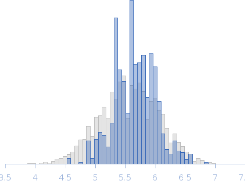
IgA peptidase (A31-I1166; active site mutant E540A) Rg histogram Rg, nm
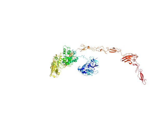
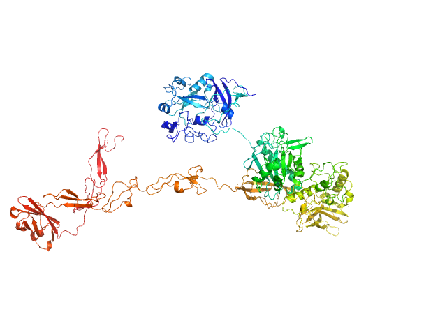
IgA protease (E540A) EOM/RANCH model

Synchrotron SAXS data from solutions of IgA peptidase in 50 mM Tris, 150 mM NaCl, pH 8 were collected on the BM29 beam line at the ESRF (Grenoble, France) using a Pilatus3 2M detector at a sample-detector distance of 2.9 m and at a wavelength of λ = 0.099 nm (I(s) vs s, where s = 4πsinθ/λ, and 2θ is the scattering angle). One solute concentration of 4.00 mg/ml was measured at 20°C. 250 successive 4 second frames were collected. The data were normalized to the intensity of the transmitted beam and radially averaged; the scattering of the solvent-blank was subtracted.

The results obtained from EOM analysis are made available in the full entry zip archive.

IgA protease (E540A) (IgAse)
Mol. type   Protein
Organism   Thomasclavelia ramosa
Olig. state   Monomer
Mon. MW   127.9 kDa
 
UniProt   Q9AES2 (31-1166)
Sequence   FASTA