Márquez-Moñino M,
Martínez Gascueña A,
Azzam T,
Persson A,
Manzanares-Gomez A,
Aguillo-Urarte M,
Brown T,
Montero-Sagarminaga A,
Lood R,
Naegeli A,
Connell S,
Sastre D,
Sundberg E,
Trastoy B,
The EMBO Journal
(2025)
DOI
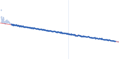
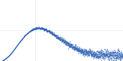
Synchrotron SAXS
data from solutions of
IgA protease (ThomasA) 323-878
in
50 mM Tris-HCl pH 7.5, 100 mM NaCl, 2% v/v glycerol, pH 7.5
were collected
on the
B21 beam line
at the Diamond Light Source storage ring
(Didcot, UK)
using a Eiger 4M detector
at a sample-detector distance of 3.7 m and
at a wavelength of λ = 0.09464 nm
(I(s) vs s, where s = 4πsinθ/λ, and 2θ is the scattering angle).
In-line size-exclusion chromatography (SEC) SAS was employed. The SEC parameters were as follows: A 55.00 μl sample
at 3.0 mg/ml was injected at a 0.07 ml/min flow rate
onto a GE Superdex 200 Increase 3.2/300 column
at 15°C.
The data were normalized to the intensity of the transmitted beam and radially averaged; the scattering of the solvent-blank was subtracted.
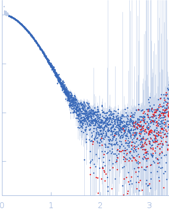 s, nm-1