|
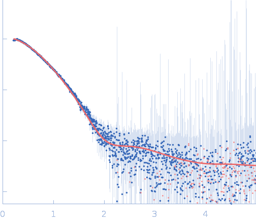
Synchrotron SAXS data from solutions of the N-terminal domain of relaxase in 20 mM Tris, 300 mM NaCl, 1 mM DTT, pH 8 were collected on the BL11-NCD beam line at ALBA (Cerdanyola del Vallès, Barcelona, Spain) using a ADSC Quantum 210r detector at a sample-detector distance of 2.6 m and at a wavelength of λ = 0.1 nm (I(s) vs s, where s = 4πsinθ/λ, and 2θ is the scattering angle). One solute concentration of 5.00 mg/ml was measured at 25°C. 20 successive 0.100 second frames were collected. The data were normalized to the intensity of the transmitted beam and radially averaged; the scattering of the solvent-blank was subtracted.
PLS20: https://doi.org/10.1093/nargab/lqab096
|
|
Relaxase
|
| Mol. type |
|
Protein |
| Organism |
|
Bacillus subtilis subsp. natto |
| Olig. state |
|
Monomer |
| Mon. MW |
|
28.5 kDa |
| |
| UniProt |
|
E9RJ23
(1-232)
|
| Sequence |
|
FASTA |
| |
|
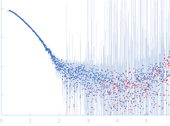 s, nm-1