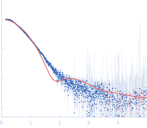
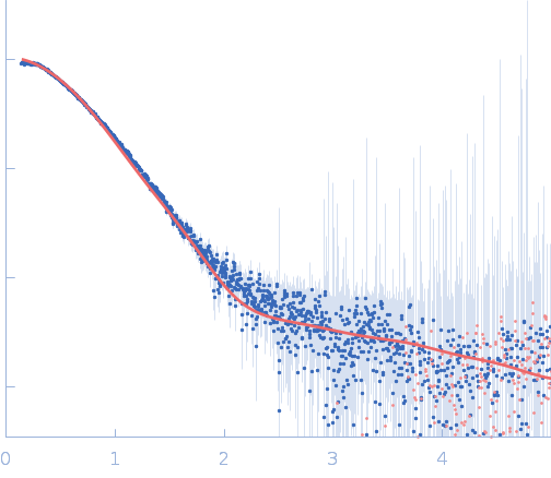
Activation and dimerization of the relaxase from the Bacillus subtilis conjugative plasmid pLS20 by DNA-induced cross-over of the catalytic residue

Isidro Crespo.

SASDX92 – N-terminal domain of relaxase from Bacillus subtilis conjugative plasmid pLS20 in complex with the core region

Relaxase
core region, single stranded
MWexperimental 49 kDa
MWexpected 69 kDa
VPorod 50 nm3
log I(s) 3.42×100 3.42×10⁻¹ 3.42×10⁻² 3.42×10⁻³
Relaxase core region, single stranded small angle scattering data  s, nm-1
ln I(s)
Relaxase core region, single stranded Guinier plot ln 3.43×100 Rg: 2.5 nm 0 (2.5 nm)-2 s2
(sRg)2I(s)/I(0)
Relaxase core region, single stranded Kratky plot 1.104 0 3 sRg
p(r)
Relaxase core region, single stranded pair distance distribution function Rg: 2.4 nm 0 Dmax: 8.2 nm

Data validation


Fits and models


log I(s)
 s, nm-1
Relaxase core region, single stranded PHENIX model

log I(s)
 s, nm-1
Relaxase core region, single stranded PYMOL model

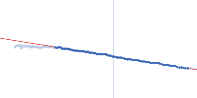
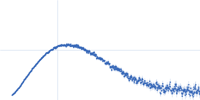
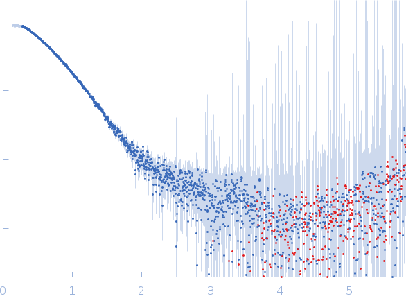
Synchrotron SAXS data from solutions of the N-terminal domain of relaxase in complex with nic sequence DNA in 20 mM Tris, 300 mM NaCl, 1 mM DTT, pH 8 were collected on the BL11-NCD beam line at ALBA (Cerdanyola del Vallès, Barcelona, Spain) using a ADSC Quantum 210r detector at a sample-detector distance of 2.6 m and at a wavelength of λ = 0.1 nm (I(s) vs s, where s = 4πsinθ/λ, and 2θ is the scattering angle). One solute concentration of 5.00 mg/ml was measured at 25°C. 20 successive 0.100 second frames were collected. The data were normalized to the intensity of the transmitted beam and radially averaged; the scattering of the solvent-blank was subtracted.

PLS20: https://doi.org/10.1093/nargab/lqab096

Relaxase
Mol. type   Protein
Organism   Bacillus subtilis subsp. natto
Olig. state   Dimer
Mon. MW   28.5 kDa
 
UniProt   E9RJ23 (1-232)
Sequence   FASTA
 
core region, single stranded (ssnic)
Mol. type   DNA
Organism   Bacillus subtilis
Olig. state   Dimer
Mon. MW   5.8 kDa
Sequence   FASTA