The role of kinase domain dimerization in EGFR activation

Zaritza Petrova.

SASDXC2 – EGFR kinase domain duplication mutant (KDD) at 1.6 mg/ml

Receptor protein-tyrosine kinase (duplication mutant)
MWI(0) 79 kDa
MWexpected 80 kDa
VPorod 135 nm3
log I(s) 1.57×10⁻¹ 1.57×10⁻² 1.57×10⁻³ 1.57×10⁻⁴
Receptor protein-tyrosine kinase (duplication mutant) small angle scattering data  s, nm-1
ln I(s)
Receptor protein-tyrosine kinase (duplication mutant) Guinier plot ln 1.57×10⁻¹ Rg: 3.9 nm 0 (3.9 nm)-2 s2
(sRg)2I(s)/I(0)
Receptor protein-tyrosine kinase (duplication mutant) Kratky plot 1.104 0 3 sRg
p(r)
Receptor protein-tyrosine kinase (duplication mutant) pair distance distribution function Rg: 4.0 nm 0 Dmax: 13 nm

Data validation


Fits and models


log I(s)
 s, nm-1
Receptor protein-tyrosine kinase (duplication mutant) DAMMIF model

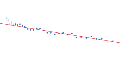
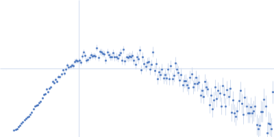
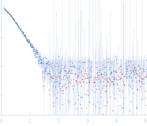
SAXS data from solutions of EGFR kinase domain duplication mutant in 20 mM HEPES, 250 mM NaCl, 250 mM KCl, pH 8 were collected on a Rigaku MicroMax-007HF instrument (Yale University, New Haven, United States) using a Rigaku PSAXS Nano detector at a sample-detector distance of 0.5 m and at a wavelength of λ = 0.1542 nm (I(s) vs s, where s = 4πsinθ/λ, and 2θ is the scattering angle). One solute concentration of 1.60 mg/ml was measured at 4°C. Three successive 7200 second frames were collected. The data were normalized to the intensity of the transmitted beam and radially averaged; the scattering of the solvent-blank was subtracted.
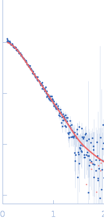
Receptor protein-tyrosine kinase (duplication mutant) (KDD)
Mol. type   Protein
Organism   Homo sapiens
Olig. state   Monomer
Mon. MW   79.6 kDa
 
UniProt   Q504U8 (651-993)
Sequence   FASTA