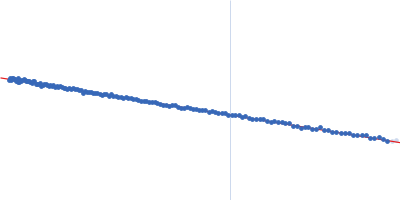
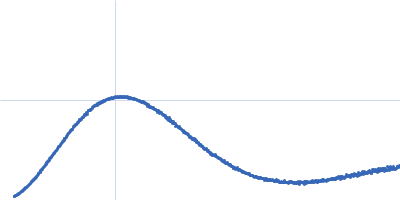
Activation of a Secondary-Messenger Receptor via Allosteric Modulation of a Dynamic Conformational Ensemble.

Söldner B, Singh H, Akoury E, Witte G, Linser R, Angew Chem Int Ed Engl :e202509394 (2025) Europe PMC

SASDXD5 – S.aureus PstA in a ligand-free state (cyclic-di-AMP receptor)

Cyclic-di-AMP receptor
MWexperimental 44 kDa
MWexpected 42 kDa
VPorod 91 nm3
log I(s) 7.42×10³ 7.42×10² 7.42×101 7.42×100
Cyclic-di-AMP receptor small angle scattering data  s, nm-1
ln I(s)
Cyclic-di-AMP receptor Guinier plot ln 7.42×10³ Rg: 2.7 nm 0 (2.7 nm)-2 s2
(sRg)2I(s)/I(0)
Cyclic-di-AMP receptor Kratky plot 1.104 0 3 sRg
p(r)
Cyclic-di-AMP receptor pair distance distribution function Rg: 2.7 nm 0 Dmax: 11 nm

Data validation


There are no models related to this curve.

Synchrotron SAXS data from solutions of S.aureus PstA in a ligand-free state (cyclic-di-AMP receptor) in 20 mM Tris, 150 mM NaCl, pH 7.5 were collected on the EMBL P12 beam line at the PETRA III storage ring (DESY; Hamburg, Germany) using a Pilatus 6M detector at a sample-detector distance of 3 m and at a wavelength of λ = 0.124402 nm (I(s) vs s, where s = 4πsinθ/λ, and 2θ is the scattering angle). Solute concentrations ranging between 2.2 and 11 mg/ml were measured at 10°C. 80 successive 0.045 second frames were collected. The data were normalized to the intensity of the transmitted beam and radially averaged; the scattering of the solvent-blank was subtracted. The low angle data collected at lower concentration were merged with the highest concentration high angle data to yield the final composite scattering curve.
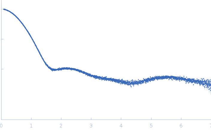
S.aureus PstA in buffer without c-di-AMP ligand. Cyclic di-AMP: https://pubchem.ncbi.nlm.nih.gov/compound/121513901

Cyclic-di-AMP receptor (PstA)
Mol. type   Protein
Organism   Staphylococcus aureus
Olig. state   Trimer
Mon. MW   14.0 kDa
 
UniProt   A0A0D1JF17 (1-109)
Sequence   FASTA