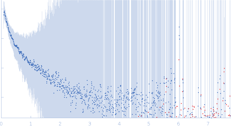
CD2AP’s Structure and Oligomerization are compromised by the K301M mutation: implications for Nephrotic syndrome

Anil Kumar Pasupulati et .al.

SASDXK6 – CD2-associated protein (CD2AP) K301M point mutant

CD2-associated protein
MWI(0) 5 kDa
MWexpected 214 kDa
log I(s) 5.85×100 5.85×10⁻¹ 5.85×10⁻² 5.85×10⁻³
CD2-associated protein small angle scattering data  s, nm-1
ln I(s)
CD2-associated protein Guinier plot ln 5.85×100 Rg: 9.6 nm 0 (9.6 nm)-2 s2
(sRg)2I(s)/I(0)
CD2-associated protein Kratky plot 1.104 0 3 sRg
p(r)
CD2-associated protein pair distance distribution function Rg: 10.7 nm 0 Dmax: 33.9 nm

Data validation


There are no models related to this curve.

SAXS data from solutions of CD2-associated protein (CD2AP) K301M point mutant in 10 mM potassium phosphate, pH 7.6 were collected on the Anton Paar SAXSpace instrument (CSIR-Central Drug Research Institute, Lucknow, India) using a Mythen2 R 1K detector at a sample-detector distance of 0.3 m and at a wavelength of λ = 0.514 nm (I(s) vs s, where s = 4πsinθ/λ, and 2θ is the scattering angle). One solute concentration of 7.00 mg/ml was measured at 10°C. Four successive 900 second frames were collected. The data were normalized to the intensity of the transmitted beam and radially averaged; the scattering of the solvent-blank was subtracted.

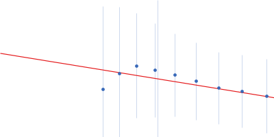
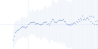
Mutant of CD2AP, First eleven points removed for display purposes. Porod volume estimation for the molecule is inconclusive.

CD2-associated protein
Mol. type   Protein
Organism   Homo sapiens
Olig. state   Trimer
Mon. MW   71.5 kDa
 
UniProt   Q9Y5K6
Sequence   FASTA