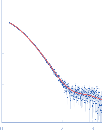
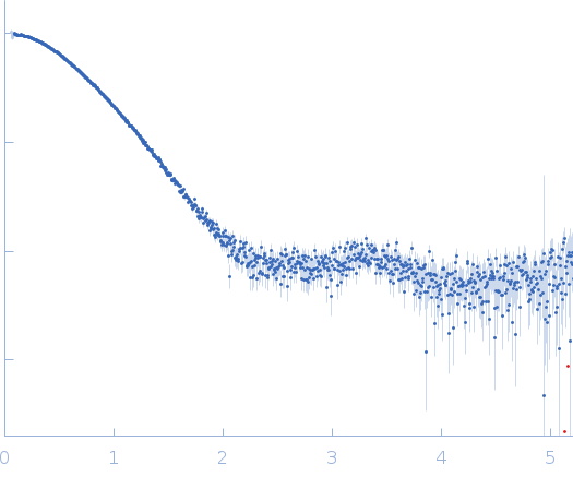
Structural and biophysical insights into RomR, MglB and MglC interactions involved in regulating cell polarity in Myxococcus xanthus

Kodesia A, Kapoor S, Thakur K, Journal of Biological Chemistry :110907 (2025) DOI

SASDXP4 – Mutual gliding motility protein C from Myxococcus xanthus

Mutual gliding motility protein C
MWexperimental 32 kDa
MWexpected 32 kDa
VPorod 49 nm3
log I(s) 4.27×101 4.27×100 4.27×10⁻¹ 4.27×10⁻²
Mutual gliding motility protein C small angle scattering data  s, nm-1
ln I(s)
Mutual gliding motility protein C Guinier plot ln 4.28×101 Rg: 2.3 nm 0 (2.3 nm)-2 s2
(sRg)2I(s)/I(0)
Mutual gliding motility protein C Kratky plot 1.104 0 3 sRg
p(r)
Mutual gliding motility protein C pair distance distribution function Rg: 2.4 nm 0 Dmax: 9.8 nm

Data validation


Fits and models


log I(s)
 s, nm-1
Mutual gliding motility protein C GASBOR model

Synchrotron SAXS data from solutions of Mutual gliding motility protein C from Myxococcus xanthus in 20 mM HEPES, 150 mM NaCl, 10% glycerol, pH 8 were collected on the BM29 beam line at the ESRF storage ring (Grenoble, France) using a Pilatus3 2M detector at a sample-detector distance of 2.8 m and at a wavelength of λ = 0.0991 nm (I(s) vs s, where s = 4πsinθ/λ, and 2θ is the scattering angle). In-line size-exclusion chromatography (SEC) SAS was employed. The SEC parameters were as follows: A 50.00 μl sample at 1 mg/ml was injected at a 0.60 ml/min flow rate onto a Agilent AdvanceBio SEC 300Å, 4.6 x 150 mm column at 4°C. 750 successive 2 second frames were collected. The data were normalized to the intensity of the transmitted beam and radially averaged; the scattering of the solvent-blank was subtracted.

Mutual gliding motility protein C (MglC)
Mol. type   Protein
Organism   Myxococcus xanthus (strain DK1622)
Olig. state   Dimer
Mon. MW   15.9 kDa
 
UniProt   Q1D0B6 (1-120)
Sequence   FASTA