DNA induces non-canonical dimerization of the farnesoid X receptor.

Khan SH, Yennawar N, Okafor CD, Nucleic Acids Res 54(4) (2026) Europe PMC

SASDXV3 – Farnesoid X receptor homo-dimer bound to inverted repeat DNA

inverted repeat DNA
Isoform 3 of Bile acid receptor
MWexperimental 95 kDa
MWexpected 94 kDa
VPorod 120 nm3
log I(s) 5.74×100 5.74×10⁻¹ 5.74×10⁻² 5.74×10⁻³
inverted repeat DNA Isoform 3 of Bile acid receptor small angle scattering data  s, nm-1
ln I(s)
inverted repeat DNA Isoform 3 of Bile acid receptor Guinier plot ln 5.75×100 Rg: 5.1 nm 0 (5.1 nm)-2 s2
(sRg)2I(s)/I(0)
inverted repeat DNA Isoform 3 of Bile acid receptor Kratky plot 1.104 0 3 sRg
p(r)
inverted repeat DNA Isoform 3 of Bile acid receptor pair distance distribution function Rg: 5.8 nm 0 Dmax: 23.2 nm

Data validation


Fits and models


log I(s)
 s, nm-1
inverted repeat DNA Isoform 3 of Bile acid receptor ALPHAFOLD model

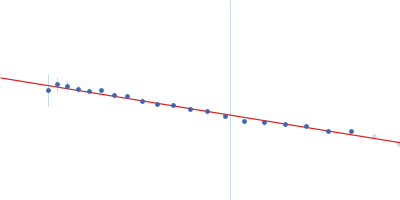
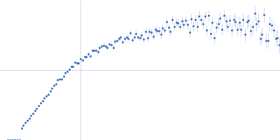
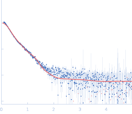
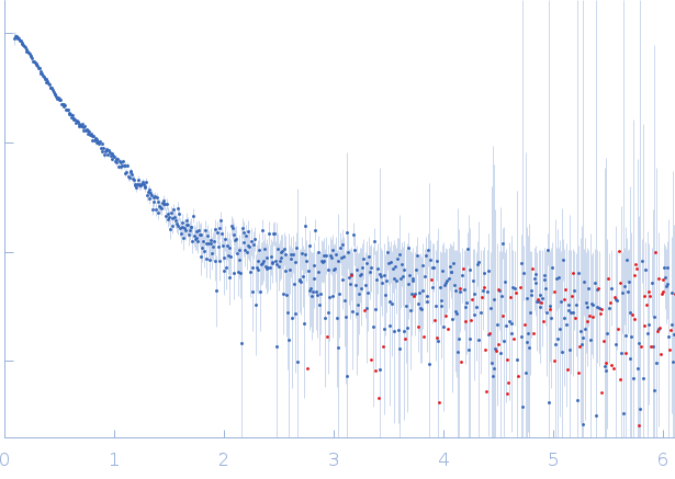
SAXS data from solutions of farnesoid X receptor homo-dimer bound to inverted repeat DNA in 50 mM HEPES, 150 mM NaCl, 5 % glycerol, 1 mM DTT, 20 µM CDCA, pH 7.4 were collected on a Rigaku BioSAXS-2000 instrument (Pennsylvania State University, University Park, PA, USA) using a Rigaku HyPix-3000 detector at a sample-detector distance of 5.0 m and at a wavelength of λ = 0.154 nm (I(s) vs s, where s = 4πsinθ/λ, and 2θ is the scattering angle). One solute concentration of 0.60 mg/ml was measured at 4°C. Six successive 600 second frames were collected. The data were normalized to the intensity of the transmitted beam and radially averaged; the scattering of the solvent-blank was subtracted.

Chenodeoxycholic acid (CDCA): https://www.ebi.ac.uk/chembl/explore/compound/CHEMBL240597

inverted repeat DNA (IR1)
Mol. type   DNA
Olig. state   Monomer
Mon. MW   11.1 kDa
Sequence   FASTA
 
Isoform 3 of Bile acid receptor (FXR)
Mol. type   Protein
Organism   Homo sapiens
Olig. state   Dimer
Mon. MW   41.3 kDa
 
UniProt   Q96RI1-1 (110-466)
Sequence   FASTA