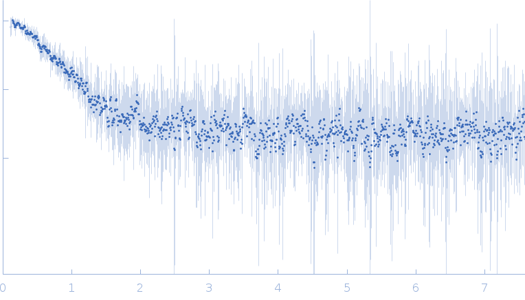
Receptor Binding Domain of SARS CoV-2

Raja Tripathi.

SASDXZ6 – Receptor Binding Domain of SARS CoV-2

Receptor Binding Domain of SARS CoV-2
MWexperimental 22 kDa
MWexpected 25 kDa
VPorod 52 nm3
log I(s) 3.10×10⁴ 3.10×10³ 3.10×10² 3.10×101
Receptor Binding Domain of SARS CoV-2 small angle scattering data  s, nm-1
ln I(s)
Receptor Binding Domain of SARS CoV-2 Guinier plot ln 3.11×10⁴ Rg: 2.9 nm 0 (2.9 nm)-2 s2
(sRg)2I(s)/I(0)
Receptor Binding Domain of SARS CoV-2 Kratky plot 1.104 0 3 sRg
p(r)
Receptor Binding Domain of SARS CoV-2 pair distance distribution function Rg: 2.3 nm 0 Dmax: 6.1 nm

Data validation


There are no models related to this curve.

SAXS data from solutions of Receptor Binding Domain of SARS CoV-2 in 50mM Tris, 200mM NaCl, pH 8 were collected on the Anton Paar SAXSpace instrument (CSIR-Central Drug Research Institute, Lucknow, India) using a Mythen2 R 1K detector at a sample-detector distance of 0.3 m and at a wavelength of λ = 0.154 nm (I(s) vs s, where s = 4πsinθ/λ, and 2θ is the scattering angle). One solute concentration of 3.00 mg/ml was measured at 10°C. Two successive 1800 second frames were collected. The data were normalized to the intensity of the transmitted beam and radially averaged; the scattering of the solvent-blank was subtracted.

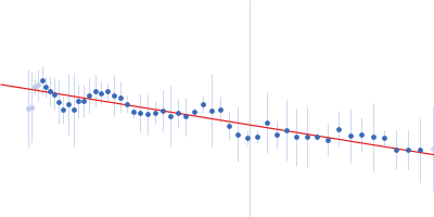
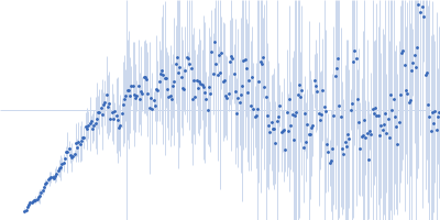
Receptor Bindind domain of SARS Cov-2 protein's SAXS data upon its purification via Size Exclusion Chromatography

Receptor Binding Domain of SARS CoV-2
Mol. type   Protein
Olig. state   Monomer
Mon. MW   25.1 kDa
 
UniProt   P0DTC2 (319-541)
Sequence   FASTA