|
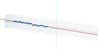
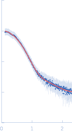
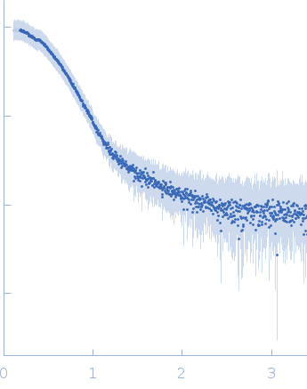
Synchrotron SAXS data from solutions of BSA in 10 mM PBS,138 mM NaCl, pH 7.25 were collected on the 4C beamline at the Pohang Accelerator Laboratory storage ring (Pohang, South Korea) using an EIGER2 X 4M detector at a sample-detector distance of 3 m and at a wavelength of λ = 0.0734 nm (I(s) vs s, where s = 4πsinθ/λ, and 2θ is the scattering angle). In-line size-exclusion chromatography (SEC) SAXS was employed. The SEC parameters were as follows: A 100.00 μl sample at 10 mg/ml was injected at a 0.1 ml/min flow rate onto a Cytiva Superdex 200 Increase 3.2/300 column at 25°C. SAXS data were collected continuously over 45 min at a flow rate of 0.1 mL/min with a 5 s integration time per frame, yielding 338 frames per run. The data were normalized to the intensity of the transmitted beam and radially averaged; the scattering of the solvent-blank was subtracted.
|
|
 s, nm-1