The phytoplasma effector, phyllogen, structurally mimics host plant MADS transcription factors.

Galien A, Hutin S, Le Hir S, Paul M, Albanese P, Kieffer-Jaquinod S, Guillotin A, Iwabuchi N, Maejima K, Nanao MH, Tully MD Hugouvieux V, Zubieta C, J Biol Chem 302(10):113449 (2026) Europe PMC

SASDY47 – Phytoplasmal effector causing phyllody 1 (Phyl1)

Phytoplasmal effector causing phyllody 1
MWexperimental 18 kDa
MWexpected 11 kDa
VPorod 51 nm3
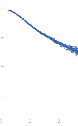
log I(s) 3.67×10⁻² 3.67×10⁻³ 3.67×10⁻⁴ 3.67×10⁻⁵
Phytoplasmal effector causing phyllody 1 small angle scattering data  s, nm-1
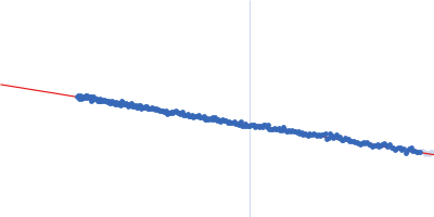
ln I(s)
Phytoplasmal effector causing phyllody 1 Guinier plot ln 3.67×10⁻² Rg: 2.2 nm 0 (2.2 nm)-2 s2
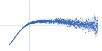
(sRg)2I(s)/I(0)
Phytoplasmal effector causing phyllody 1 Kratky plot 1.104 0 √3 sRg
p(r)
Phytoplasmal effector causing phyllody 1 pair distance distribution function Rg: 2.1 nm 0 Dmax: 6.6 nm

Data validation


There are no models related to this curve.

Synchrotron SAXS data from solutions of Phytoplasmal effector causing phyllody 1 (Phyl1) in 50 mM Tris, 300 mM NaCl, 1 mM TCEP, pH 8 were collected on the B21 beam line at the Diamond Light Source storage ring (Didcot, UK) using a Eiger 4M detector at a sample-detector distance of 3.7 m and at a wavelength of λ = 0.1 nm (I(s) vs s, where s = 4πsinθ/λ, and 2θ is the scattering angle). One solute concentration of 14.30 mg/ml was measured at 20°C. 620 successive 1 second frames were collected. The data were normalized to the intensity of the transmitted beam and radially averaged; the scattering of the solvent-blank was subtracted.

Collected via SEC-SAXS, helix with flexible linker

Phytoplasmal effector causing phyllody 1 (PHYL1)
Mol. type   Protein
Organism   Onion yellows phytoplasma OY-W
Olig. state   Monomer
Mon. MW   10.7 kDa
 
UniProt   X5IFG3 (35-125)
Sequence   FASTA