Dmax unknown – experimental data range validation not possible.
There are no models related to this curve.
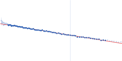
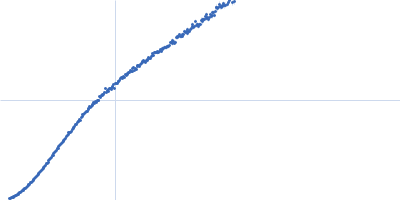
Synchrotron SAXS
data from solutions of
p53 proline-rich domain of Cellular tumor antigen p53 with (P82L) with 10 mM NaCl and neutral pH
in
20 mM Tris, 10 mM NaCl,, pH 7
were collected
on the
BM29 beam line
at the ESRF storage ring
(Grenoble, France)
using a Pilatus3 2M detector
at a sample-detector distance of 2.9 m and
at a wavelength of λ = 0.0992 nm
(I(s) vs s, where s = 4πsinθ/λ, and 2θ is the scattering angle).
One solute concentration of 8.05 mg/ml was measured
at 20°C.
10 successive
1 second frames were collected.
The data were normalized to the intensity of the transmitted beam and radially averaged; the scattering of the solvent-blank was subtracted.
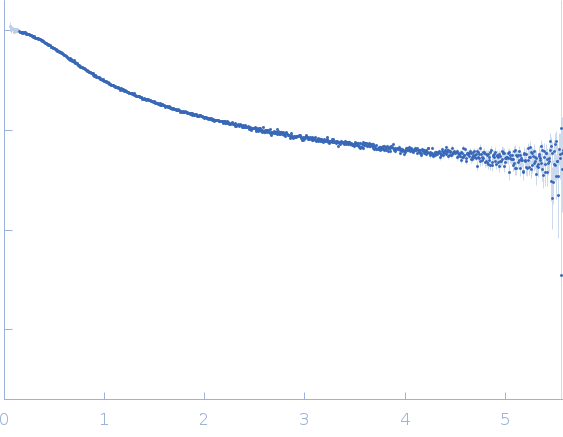 s, nm-1