Synchrotron SAXS
data from solutions of
Ferric anguibactin-binding protein, apo (FatB)
in
20mM Tris-HCl, 100 mM NaCl, pH 8
were collected
on the
4C beam line
at the Pohang Accelerator Laboratory storage ring
(Pohang, South Korea)
using a Dectris / EIGER2 X 4M detector
at a sample-detector distance of 3 m and
at a wavelength of λ = 0.0734 nm
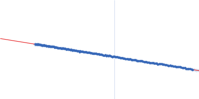
(I(s) vs s, where s = 4πsinθ/λ, and 2θ is the scattering angle).
One solute concentration of 3.00 mg/ml was measured
at 4°C.
50 successive
10 second frames were collected.
The data were normalized to the intensity of the transmitted beam and radially averaged; the scattering of the solvent-blank was subtracted.
SAXS data of apo-FatB were collected at two sample-to-detector distances (3 m and 1 m). The 3 m setting covered the low-q region (q ≈ 0.025 to 0.125 Å-1), and the 1 m setting covered the higher-q region (q ≈ 0.125 to 0.6 Å-1). The datasets were scaled and checked for agreement in the overlap region before merging into a single profile.
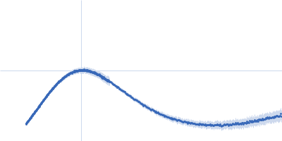
 s, nm-1