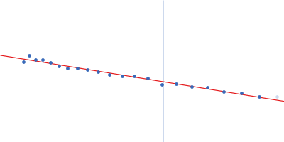
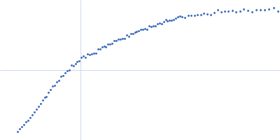
Dmax unknown – experimental data range validation not possible.
There are no models related to this curve.
Synchrotron SAXS
data from solutions of
Poly-cytosine rC30 RNA in 100 mM NaCl
in
100 mM NaCl, 1 mM MOPS, 20 µM EDTA, pH 7.1
were collected
on the
ID7A1 BioSAXS / HP-Bio Beamline beam line
at the Cornell High Energy Synchrotron Source (CHESS) storage ring
(Ithaca, NY, USA)
using a Eiger 4M detector
at a sample-detector distance of 1.8 m and
at a wavelength of λ = 0.1094 nm
(I(s) vs s, where s = 4πsinθ/λ, and 2θ is the scattering angle).
One solute concentration of 0.90 mg/ml was measured
at 25°C.
100 successive
1 second frames were collected.
The data were normalized to the intensity of the transmitted beam and radially averaged; the scattering of the solvent-blank was subtracted.

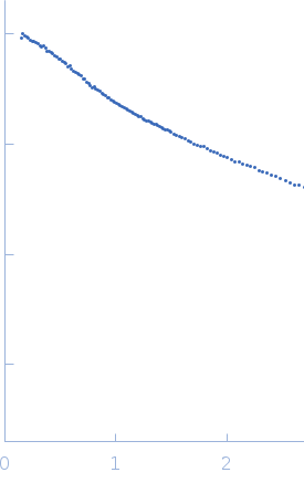 s, nm-1