| MWexperimental | 4 | kDa |
| MWexpected | 4 | kDa |
| VPorod | 7 | nm3 |
|
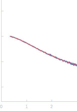
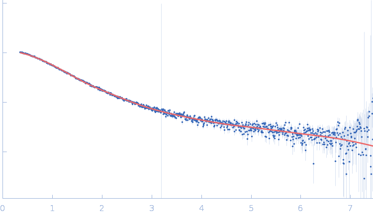
log I(s)
2.86×101
2.86×100
2.86×10⁻¹
2.86×10⁻²
|
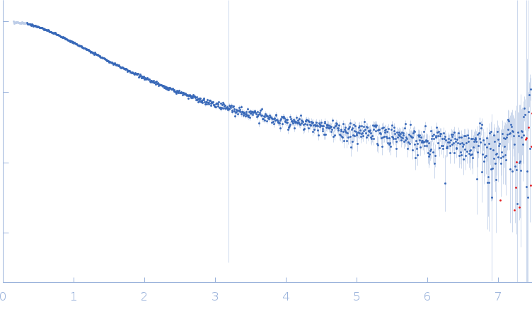 s, nm-1
|
|
|
|

|
|
|
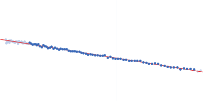
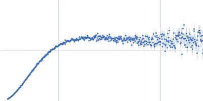
Synchrotron SAXS
data from solutions of
Histatin 3 peptide
in
20 mM HEPES, 150 mM NaCl, pH 7.4
were collected
on the
BL4-2 beam line
at the Stanford Synchrotron Radiation Lightsource (SSRL) storage ring
(Menlo Park, CA, USA)
using a Pilatus3 X 1M detector
at a sample-detector distance of 1.2 m and
at a wavelength of λ = 0.1127 nm
(I(s) vs s, where s = 4πsinθ/λ, and 2θ is the scattering angle).
One solute concentration of 10.00 mg/ml was measured
at 15°C.
16 successive
1 second frames were collected.
The data were normalized to the intensity of the transmitted beam and radially averaged; the scattering of the solvent-blank was subtracted.
|
|
|||||||||||||||||||||||||||