Dmax unknown – experimental data range validation not possible.
There are no models related to this curve.
SAXS
data from solutions of
endo-β-1,4-xylanase (AcXyn30B_12) from Acetivibrio clariflavus
in
50 mM Sodium Phosphate, pH 7.5
were collected
on the
Anton Paar SAXSpace instrument (Medical University of Graz, Graz, Austria)
using a Roper Scientific PI-SCX:4300 detector
at a sample-detector distance of 1 m and
at a wavelength of λ = 1.54 nm
(I(s) vs s, where s = 4πsinθ/λ, and 2θ is the scattering angle).
One solute concentration of 3.00 mg/ml was measured
at 10°C.
The data were normalized to the intensity of the transmitted beam and radially averaged; the scattering of the solvent-blank was subtracted.
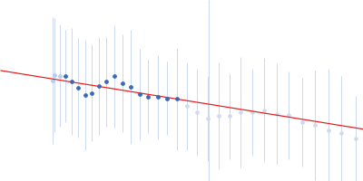
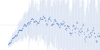
CAUTION: The reported experimental uncertainties appear inconsistent with the variation in the data, preventing a clear identification of the Guinier region. Derived parameters should be interpreted with caution
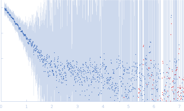 s, nm-1