Okuda A
Inoue R,
Kurokawa M,
Martel A,
Porcar L,
Osaki R,
Fukuzawa K,
Weiss K,
Pingali S,
Urade R,
Sugiyama M,
Journal of Applied Crystallography
59(4):1270-1281
(2026)
DOI
SASDZS8 – Segmental deuterated Protein disulfide-isomerase A3 comprising three point mutations (D122N/I124L/R363N/Y364G; ER-60 h(a)-pd(bb’)-h(a’))
Dmax unknown – experimental data range validation not possible.
There are no models related to this curve.
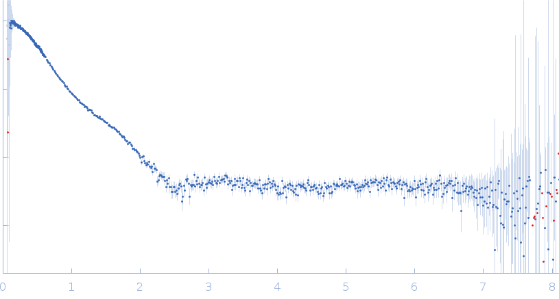
SAXS
data from solutions of
Segmental deuterated Protein disulfide-isomerase A3 comprising three point mutations (D122N/I124L/R363N/Y364G; ER-60 h(a)-pd(bb’)-h(a’))
in
20 mM Tris-HCl buffer, 150 mM NaCl, 1mM CaCl2, pH 8
were collected
on the
Rigaku NANOPIX instrument (KURNS, Osaka, Japan)
using a HyPix-6000 detector
at a sample-detector distance of 1.3 m and
at a wavelength of λ = 1.54182 nm
(I(s) vs s, where s = 4πsinθ/λ, and 2θ is the scattering angle).
Solute concentrations ranging between 4 and 4 mg/ml were measured
at 25°C.
72 successive
300 second frames were collected.
The data were normalized to the intensity of the transmitted beam and radially averaged; the scattering of the solvent-blank was subtracted.
The low angle data collected at lower concentration were merged with the highest concentration high angle data to yield the final composite scattering curve.
Two sample-to-detector distance (SDD) conditions, 1330 mm and 300 mm, were used to cover a q range (0.01-0.70 Å-1). Scattering profiles were recorded as 300 s frames, and 72 and 120 frames merged for low q (0.01-0.06 Å-1) and high q (0.06-0.70 Å-1) setups, respectively. We applied the AUC-SAS method to remove aggregation components from the profile.

 s, nm-1