Structural Analysis of the Menangle Virus P Protein Reveals a Soft Boundary between Ordered and Disordered Regions
Webby M,
Herr N,
Bulloch E,
Schmitz M,
Keown J,
Goldstone D,
Kingston R
Viruses
13(9):1737
(2021 Aug 31)
doi: 10.3390/v13091737
|
Submitted to SASBDB: 2021 Jul 5
Published in SASBDB:
|
|
|
|
|
|
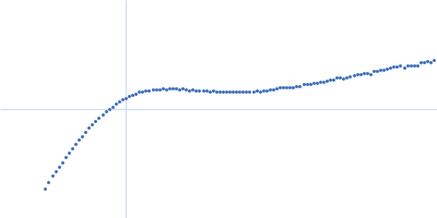
| Sample: |
Phosphoprotein tetramer, 79 kDa Menangle virus protein
|
| Buffer: |
12.5 mM MOPS/KOH pH 7.0, 250 mM NaCl, pH: 7 |
| Experiment: |
SAXS
data collected at SAXS/WAXS, Australian Synchrotron on 2016 Nov 16
|
|
| RgGuinier |
6.3 |
nm |
| Dmax |
23.4 |
nm |
| VolumePorod |
524 |
nm3 |
|
|
|
|
|
|
|
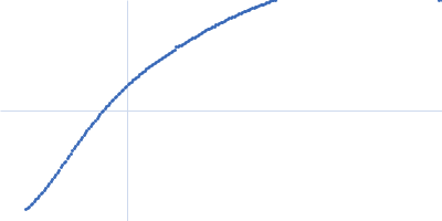
| Sample: |
Phosphoprotein monomer, 13 kDa Menangle virus protein
|
| Buffer: |
12.5 mM MOPS/KOH pH 7.0, 150 mM NaCl, pH: 7 |
| Experiment: |
SAXS
data collected at SAXS/WAXS, Australian Synchrotron on 2016 Aug 17
|
|
| RgGuinier |
3.1 |
nm |
| Dmax |
12.2 |
nm |
| VolumePorod |
20 |
nm3 |
|
|
|
|
|
|
|
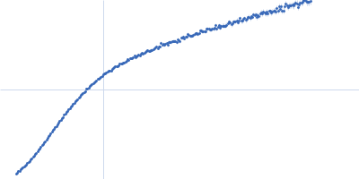
| Sample: |
Phosphoprotein monomer, 6 kDa Menangle virus protein
|
| Buffer: |
12.5 mM Tris/HCl pH 8.5, 150 mM NaCl, pH: 8.5 |
| Experiment: |
SAXS
data collected at SAXS/WAXS, Australian Synchrotron on 2017 Aug 15
|
|
| RgGuinier |
2.5 |
nm |
| Dmax |
10.3 |
nm |
| VolumePorod |
12 |
nm3 |
|
|