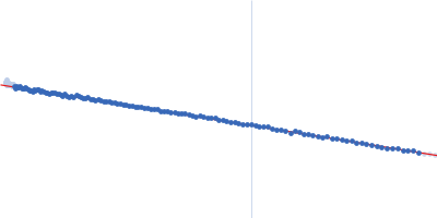
Synchrotron Size-exclusion coupled to Small angle X-ray scattering (SEC-SAXS) data from non-methylated Virulence factor family protein (VirJD1) from Brucella abortus in 50 mM Tris pH 8.0, 200 mM NaCl, 5% v/v glycerol were collected on the SWING beamline at the SOLEIL storage ring (Saint-Aubin, France) using an Eiger 4M detector at a sample-detector distance of 2 m and at a wavelength of λ = 0.1033 nm (I(s) vs s, where s = 4πsinθ/λ, and 2θ is the scattering angle). The SEC parameters were as follows: Fifty μl of protein at 15 mg/mL were loaded onto a size-exclusion column (S200 increase 5/150) at a 0.20 ml/min flow rate at 20°C. Frames were recorded every second for the duration of the size-exclusion run. Buffer subtraction was performed by averaging 100 buffer frames and 16 successive sample frames. The data were normalized to the intensity of the transmitted beam and radially averaged; the scattering of the solvent-blank was subtracted.
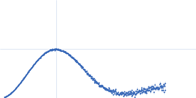
 s, nm-1
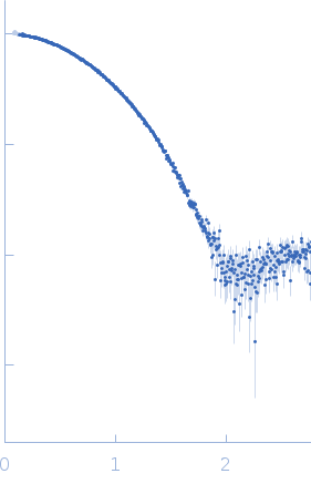