A structural basis for chaperone repression of stress signaling from the endoplasmic reticulum.

Neidhardt L, Tung J, Kuchersky M, Milczarek J, Kargas V, Stott K, Rosenzweig R, Ron D, Yan Y, Mol Cell (2025) Europe PMC

SASDWU6 – Human anterior gradient protein 2 (AGR2)

Anterior gradient protein 2 homolog
MWexperimental 24 kDa
MWexpected 30 kDa
VPorod 30 nm3
log I(s) 1.77×10⁻¹ 1.77×10⁻² 1.77×10⁻³ 1.77×10⁻⁴
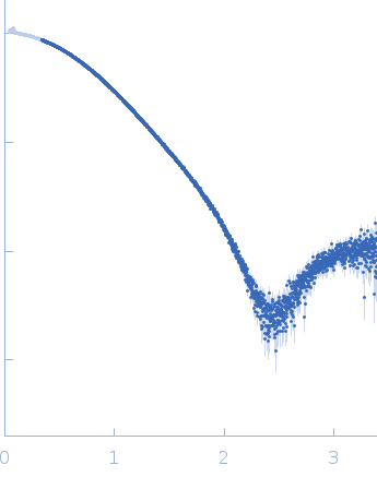
Anterior gradient protein 2 homolog small angle scattering data  s, nm-1
ln I(s)
Anterior gradient protein 2 homolog Guinier plot ln 1.78×10⁻¹ Rg: 2.0 nm 0 (2.0 nm)-2 s2
(sRg)2I(s)/I(0)
Anterior gradient protein 2 homolog Kratky plot 1.104 0 3 sRg
p(r)
Anterior gradient protein 2 homolog pair distance distribution function Rg: 2.0 nm 0 Dmax: 6.3 nm

Data validation


Fits and models


log I(s)
 s, nm-1
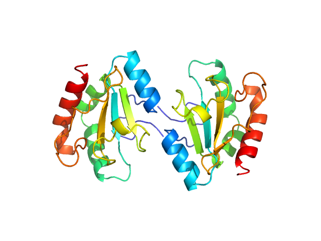
Anterior gradient protein 2 homolog MULTIFOXS model

Synchrotron SAXS data from solutions of anterior gradient protein 2 (AGR2) in 20 mM Tris pH7.4, 150 mM NaCl, 1% glycerol, 0.2 mM TCEP, were collected on the B21 beam line at the Diamond Light Source (Didcot, UK) using a Eiger 4M detector at a sample-detector distance of 3.7 m and at a wavelength of λ = 0.09464 nm (I(s) vs s, where s = 4πsinθ/λ, and 2θ is the scattering angle). In-line size-exclusion chromatography (SEC) SAS was employed. The SEC parameters were as follows: A 35.00 μl sample at 46 mg/ml was injected at a 0.06 ml/min flow rate onto a GE Superdex 75 Increase 3.2/300 column at 15°C. 30 successive 1 second frames were collected through the sample elution peak (from a total of 600 frames collected throughout the entire SEC run). The data were normalized to the intensity of the transmitted beam and radially averaged; the scattering of the solvent-blank was subtracted.

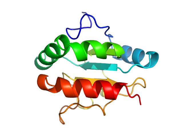
Anterior gradient protein 2 homolog (AGR2)
Mol. type   Protein
Organism   Homo sapiens
Olig. state   Dimer
Mon. MW   15.1 kDa
 
UniProt   O95994 (41-171)
Sequence   FASTA