The structure of the apolipoprotein A-I monomer provides insights into its oligomerisation and lipid-binding mechanisms

Tou H, Rosenes Z, Khandokar Y, Zlatic C, Metcalfe R, Mok Y, Morton C, Gooley P, Griffin M, Journal of Molecular Biology :169394 (2025) DOI

SASDUB9 – Apolipoprotein A-I monomer, C-terminally truncated, SAXS construct G26R mutant

Apolipoprotein A-I (G50R)
MWexperimental 17 kDa
MWexpected 22 kDa
VPorod 28 nm3
log I(s) 1.14×10⁻² 1.14×10⁻³ 1.14×10⁻⁴ 1.14×10⁻⁵
Apolipoprotein A-I (G50R) small angle scattering data  s, nm-1
ln I(s)
Apolipoprotein A-I (G50R) Guinier plot ln 1.15×10⁻² Rg: 2.3 nm 0 (2.3 nm)-2 s2
(sRg)2I(s)/I(0)
Apolipoprotein A-I (G50R) Kratky plot 1.104 0 3 sRg
p(r)
Apolipoprotein A-I (G50R) pair distance distribution function Rg: 2.4 nm 0 Dmax: 7.9 nm

Data validation


There are no models related to this curve.

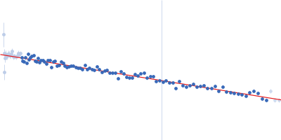
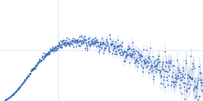
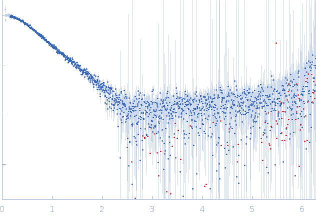
Synchrotron SAXS data from solutions of C-terminally truncated Apolipoprotein A-I (G26R) in 20 mM Tris, 150 mM NaCl, 0.1% sodium azide, pH 7.4 were collected on the SAXS/WAXS beam line at the Australian Synchrotron (Melbourne, Australia) using a Pilatus3 S 2M detector at a wavelength of λ = 0.1078 nm (I(s) vs s, where s = 4πsinθ/λ, and 2θ is the scattering angle). One solute concentration of 5.00 mg/ml was measured at 20°C. 13 successive 1 second frames were collected. The data were normalized to the intensity of the transmitted beam and radially averaged; the scattering of the solvent-blank was subtracted.

The point mutation occurs at position 26 (G26R) in the protein construct used for SAXS. Relative to the canonical amino acid sequence of the full length protein listed in UniProt - that includes an N-terminal signal peptide - the amino acid substitution referred to here occurs at position 50 (UniProt P02647; G50R). Sample detector distance: UNKNOWN.

Apolipoprotein A-I (G50R) (G50R apoA-I)
Mol. type   Protein
Organism   Homo sapiens
Olig. state   Monomer
Mon. MW   21.7 kDa
 
UniProt   P02647 (25-208)
Sequence   FASTA