|
Synchrotron SAXS
data from solutions of
Native state of monomeric full-length glycoside hydrolase family18 chitodextrinase from Vibrio cholerae
in
100mM HEPES, 150mM NaCl, pH 7.5
were collected
on the
TPS13A beam line
at the NSRRC storage ring
(Hsinchu, Taiwan)
using a Eiger X 1M & 9M detector
at a sample-detector distance of 2.5 m and
at a wavelength of λ = 0.0827 nm
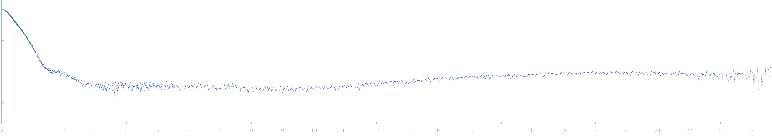
(I(s) vs s, where s = 4πsinθ/λ, and 2θ is the scattering angle).
One solute concentration of 5.00 mg/ml was measured
at 10°C.
240 successive
2 second frames were collected.
The data were normalized to the intensity of the transmitted beam and radially averaged; the scattering of the solvent-blank was subtracted.
The SAS profile obtained by SEC-SAXS connected with Superdex200-increase column size 5x150 mm (Cytiva), flow rate 0.15 mL/min. The 100 µL of 5 mg/mL protein was injected.
|
|
Chitodextrinase
(ChDex)
|
| Mol. type |
|
Protein |
| Organism |
|
Vibrio cholerae |
| Olig. state |
|
Monomer |
| Mon. MW |
|
108.5 kDa |
| |
| UniProt |
|
Q9KLP3
|
| Sequence |
|
FASTA |
| |
|
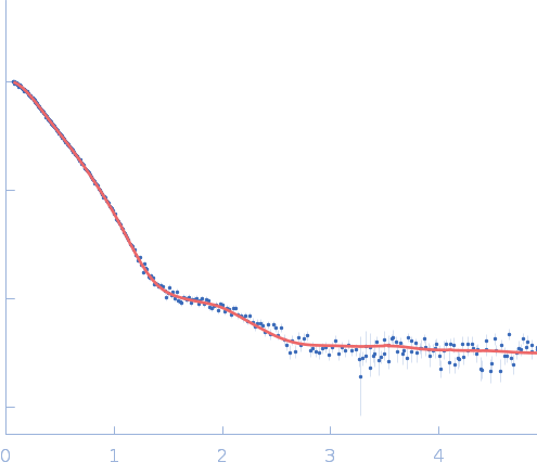
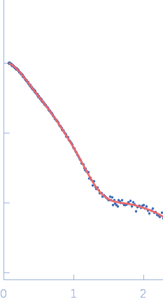
 s, nm-1
 Rg, nm