|
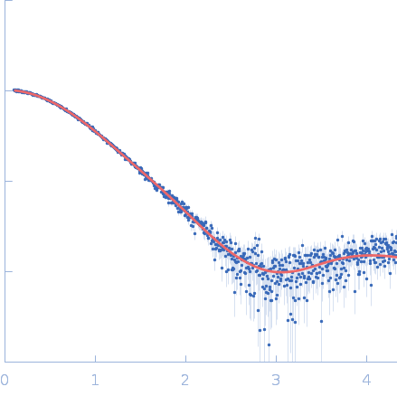
Synchrotron SAXS
data from solutions of
Influenza A virus Nuclear export protein NEP/NS2
in
20 mM HEPES, 300 mM NaCl, pH 7.5
were collected
on the
SWING beam line
at the SOLEIL storage ring
(Saint-Aubin, France)
using a Eiger 4M detector
at a sample-detector distance of 2 m and
at a wavelength of λ = 0.1033 nm
(I(s) vs s, where s = 4πsinθ/λ, and 2θ is the scattering angle).
In-line size-exclusion chromatography (SEC) SAS was employed. The SEC parameters were as follows: A 15.00 μl sample
at 6 mg/ml was injected at a 0.30 ml/min flow rate
onto a GE Superdex 75 Increase 5/150 column
at 20°C.
400 successive
1 second frames were collected.
The data were normalized to the intensity of the transmitted beam and radially averaged; the scattering of the solvent-blank was subtracted.
|
|
Nuclear export protein
(NEP)
|
| Mol. type |
|
Protein |
| Organism |
|
Influenza A virus (strain A/Wilson-Smith/1933 H1N1) (Influenza A virus (strain A/WS/1933 H1N1)) |
| Olig. state |
|
Monomer |
| Mon. MW |
|
14.3 kDa |
| |
| UniProt |
|
Q89733
|
| Sequence |
|
FASTA |
| |
|
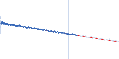
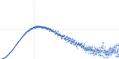
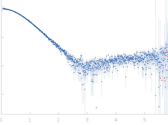 s, nm-1