Crystal structure and molecular dynamics of human POLDIP2, a multifaceted adaptor protein in metabolism and genome stability.

Kulik AA, Maruszczak KK, Thomas DC, Nabi-Aldridge NLA, Carr M, Bingham RJ, Cooper CDO, Protein Sci (2021) Europe PMC

SASDK76 – Polymerase delta-interacting protein 2 (POLDIP2, amino acids 51-368)

Polymerase delta-interacting protein 2
MWexperimental 37 kDa
MWexpected 37 kDa
VPorod 56 nm3
log I(s) 1.52×101 1.52×100 1.52×10⁻¹ 1.52×10⁻²
Polymerase delta-interacting protein 2 small angle scattering data  s, nm-1
ln I(s)
Polymerase delta-interacting protein 2 Guinier plot ln 1.53×101 Rg: 2.2 nm 0 (2.2 nm)-2 s2
(sRg)2I(s)/I(0)
Polymerase delta-interacting protein 2 Kratky plot 1.104 0 3 sRg
p(r)
Polymerase delta-interacting protein 2 pair distance distribution function Rg: 2.3 nm 0 Dmax: 7.2 nm

Data validation


There are no models related to this curve.

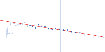
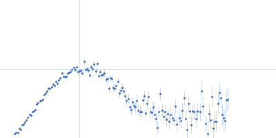
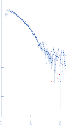
SAXS data from solutions of POLDIP2 (amino acids 51-368) in 20 mM HEPES pH 7.5, 500 mM NaCl, 5% (v/v) glycerol, were collected using a Bruker Nanostar instrument (University of Huddersfield, West Yorkshire, United Kingdom) equipped with a VÅNTEC-2000 detector at a sample-detector distance of 1.1 m and at a wavelength of λ = 0.154 nm (I(s) vs s, where s = 4πsinθ/λ, and 2θ is the scattering angle). One solute concentration of 3.40 mg/ml was measured at 20°C. 10 successive 1800 second frames were collected. The data were normalized to the intensity of the transmitted beam and radially averaged; the scattering of the solvent-blank was subtracted.

NOTE: The protein sequence has an additional two N-terminal amino acids (SM) prior to amino acid 51 of the POLDIP2 UniProt sequence.

Polymerase delta-interacting protein 2 (POLDIP2)
Mol. type   Protein
Organism   Homo sapiens
Olig. state   Monomer
Mon. MW   37.0 kDa
 
UniProt   Q9Y2S7 (51-368)
Sequence   FASTA