|
|
|
|
|
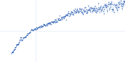
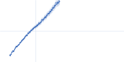
| Sample: |
Collagenase ColG (E656D, N659T, G836V, D837Y) monomer, 114 kDa Hathewaya histolytica protein
|
| Buffer: |
10 mM HEPES, 100 mM NaCl, 1 mM CaCl2, EDTA, pH: 7.5 |
| Experiment: |
SAXS
data collected at 12.3.1 (SIBYLS), Advanced Light Source (ALS) on 2022 Oct 28
|
Collagenase G
Cody Brazel
|
| RgGuinier |
4.8 |
nm |
| Dmax |
18.1 |
nm |
| VolumePorod |
182 |
nm3 |
|
|
|
|
|
|
|
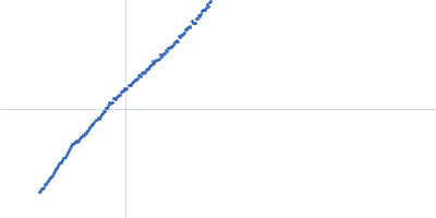
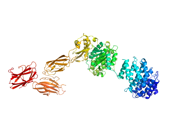
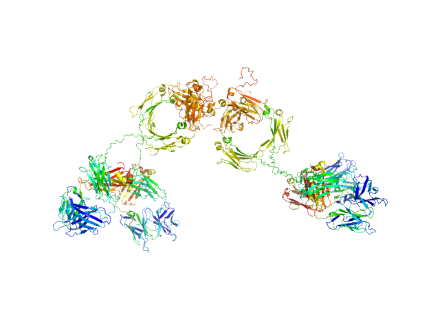
| Sample: |
Human immunoglobulin IgA1 dimer, 320 kDa protein
|
| Buffer: |
8.2 mM Na2HPO4, 1.5 mM KH2PO4, 137 mM NaCl, 2.6 mM KCl (PBS), pH: 7.4 |
| Experiment: |
SAXS
data collected at BM29, ESRF on 2014 Sep 7
|
Atomistic scattering modelling of the solution structure of human dimeric IgA1 reveals a structural and mechanistic basis for IgA nephropathy.
J Biol Chem :113156 (2026)
Bhatt JS, Yeo SC, Ireland SM, Ben-Younis A, Gor J, Molyneux K, Barratt J, Perkins SJ
|
| RgGuinier |
8.7 |
nm |
| Dmax |
35.0 |
nm |
|
|
|
|
|
|
|
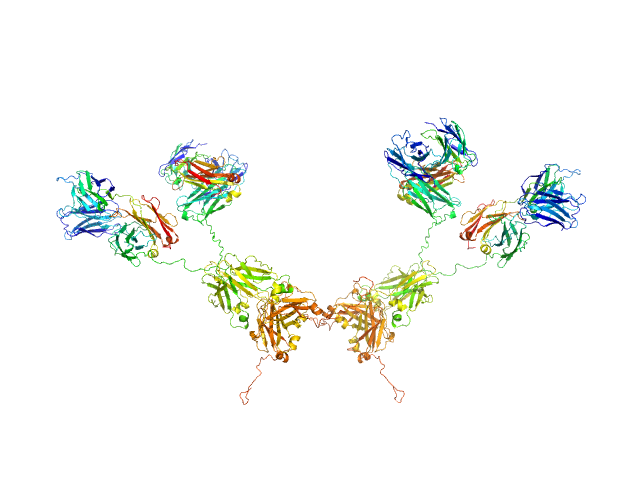
| Sample: |
Human immunoglobulin IgA1 dimer, 320 kDa protein
|
| Buffer: |
PBS buffer, 137 mM NaCl, 8.2 mM Na2HPO4, 2.6 mM KCl, 1.5 mM KH2PO4, pH: 7.4 |
| Experiment: |
SAXS
data collected at BM29, ESRF on 2014 Sep 7
|
Atomistic scattering modelling of the solution structure of human dimeric IgA1 reveals a structural and mechanistic basis for IgA nephropathy.
J Biol Chem :113156 (2026)
Bhatt JS, Yeo SC, Ireland SM, Ben-Younis A, Gor J, Molyneux K, Barratt J, Perkins SJ
|
| RgGuinier |
8.6 |
nm |
| Dmax |
35.0 |
nm |
|
|
|
|
|
|
|
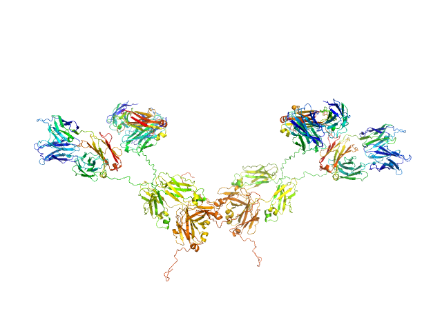
| Sample: |
Human immunoglobulin IgA1 dimer, 320 kDa protein
|
| Buffer: |
PBS buffer, 137 mM NaCl, 8.2 mM Na2HPO4, 2.6 mM KCl, 1.5 mM KH2PO4, pH: 7.4 |
| Experiment: |
SAXS
data collected at BM29, ESRF on 2014 Sep 7
|
Atomistic scattering modelling of the solution structure of human dimeric IgA1 reveals a structural and mechanistic basis for IgA nephropathy.
J Biol Chem :113156 (2026)
Bhatt JS, Yeo SC, Ireland SM, Ben-Younis A, Gor J, Molyneux K, Barratt J, Perkins SJ
|
| RgGuinier |
8.4 |
nm |
| Dmax |
35.0 |
nm |
|
|
|
|
|
|
|
| Sample: |
Human immunoglobulin IgA1 dimer, 320 kDa protein
|
| Buffer: |
PBS buffer, 137 mM NaCl, 8.2 mM Na2HPO4, 2.6 mM KCl, 1.5 mM KH2PO4, pH: 7.4 |
| Experiment: |
SAXS
data collected at BM29, ESRF on 2014 Sep 7
|
Atomistic scattering modelling of the solution structure of human dimeric IgA1 reveals a structural and mechanistic basis for IgA nephropathy.
J Biol Chem :113156 (2026)
Bhatt JS, Yeo SC, Ireland SM, Ben-Younis A, Gor J, Molyneux K, Barratt J, Perkins SJ
|
| RgGuinier |
8.5 |
nm |
| Dmax |
35.0 |
nm |
|
|
|
|
|
|
|
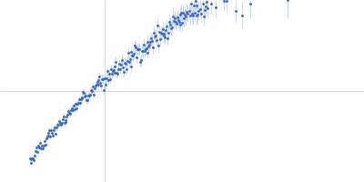
| Sample: |
Alpha-actinin-2 dimer, 208 kDa Homo sapiens protein
|
| Buffer: |
20 mM HEPES, 150 mM NaCl, pH: 8 |
| Experiment: |
SAXS
data collected at B21, Diamond Light Source on 2024 May 13
|
ACTN2 HCM-linked Missense Variants
Maya Noureddine
|
| RgGuinier |
12.1 |
nm |
| Dmax |
40.6 |
nm |
|
|
|
|
|
|
|
| Sample: |
Alpha-actinin-2 dimer, 208 kDa Homo sapiens protein
|
| Buffer: |
20 mM HEPES, 150 mM NaCl, pH: 8 |
| Experiment: |
SAXS
data collected at B21, Diamond Light Source on 2024 May 13
|
ACTN2 HCM-linked Missense Variants
Maya Noureddine
|
| RgGuinier |
11.9 |
nm |
| Dmax |
36.2 |
nm |
|
|
|
|
|
|
|
| Sample: |
Alpha-actinin-2 dimer, 208 kDa Homo sapiens protein
|
| Buffer: |
20 mM HEPES, 150 mM NaCl, pH: 8 |
| Experiment: |
SAXS
data collected at B21, Diamond Light Source on 2024 May 13
|
ACTN2 HCM-linked Missense Variants
Maya Noureddine
|
| RgGuinier |
10.9 |
nm |
| Dmax |
37.0 |
nm |
|
|
|
|
|
|
|
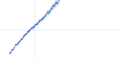
| Sample: |
Alpha-actinin-2 dimer, 208 kDa Homo sapiens protein
|
| Buffer: |
20 mM HEPES, 150 mM NaCl, pH: 8 |
| Experiment: |
SAXS
data collected at B21, Diamond Light Source on 2024 May 13
|
ACTN2 HCM-linked Missense Variants
Maya Noureddine
|
| RgGuinier |
10.3 |
nm |
| Dmax |
35.3 |
nm |
|
|
|
|
|
|
|
| Sample: |
Alpha-actinin-2 dimer, 208 kDa Homo sapiens protein
|
| Buffer: |
20 mM HEPES, 150 mM NaCl, pH: 8 |
| Experiment: |
SAXS
data collected at B21, Diamond Light Source on 2024 May 13
|
ACTN2 HCM-linked Missense Variants
Maya Noureddine
|
| RgGuinier |
10.3 |
nm |
| Dmax |
37.0 |
nm |
|
|
 experimental SAS data")