|
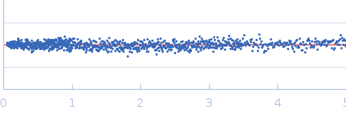
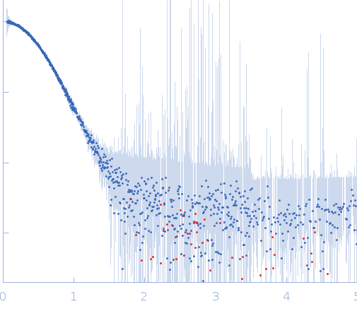
Synchrotron SAXS
data from solutions of
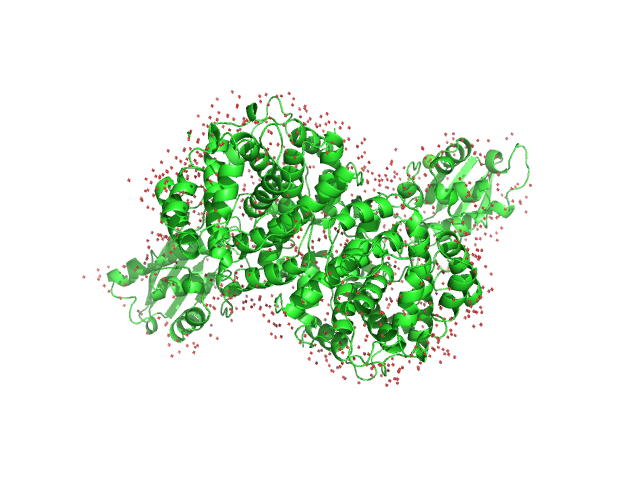
Cis-aconitate decarboxylase ACOD1 ACOD1 Arg273Ser mutant
in
20 mM HEPES , 150 mM NaCl, 0.5 mM TCEP, and 2% (v/v) glycerol, pH 7.5
were collected
on the
12-ID-B beam line
at the Advanced Photon Source (APS), Argonne National Laboratory storage ring
(Lemont, IL, USA)
using a Eiger2 S detector
at a sample-detector distance of 2 m and
at a wavelength of λ = 0.0932 nm
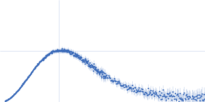
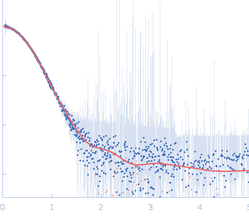
(I(s) vs s, where s = 4πsinθ/λ, and 2θ is the scattering angle).
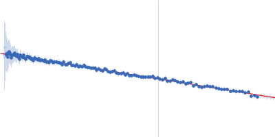
In-line size-exclusion chromatography (SEC) SAS was employed. The SEC parameters were as follows: A 100.00 μl sample
at 6.1 mg/ml was injected at a 0.30 ml/min flow rate
onto a GE Superdex 200 Increase 5/150 column
at 20°C.
694 successive
0.300 second frames were collected.
The data were normalized to the intensity of the transmitted beam and radially averaged; the scattering of the solvent-blank was subtracted.
|
|

 s, nm-1