Solution structures of glycosaminoglycan-bound CXCL8 complexes determined by small-angle X-ray scattering (SAXS).

White MA Mahler BP, Joseph PRB, Nagarajan B, Wang X, Desai UR, Rajarathnam K, Biochem J 483(10):1861-1876 (2026) Europe PMC

SASD2S2 – Interleukin-8-Heparin sulfate (CXCL8:Heparin)

INTERLEUKIN 8
Heparin 8-mer
MWexperimental 28 kDa
MWexpected 18 kDa
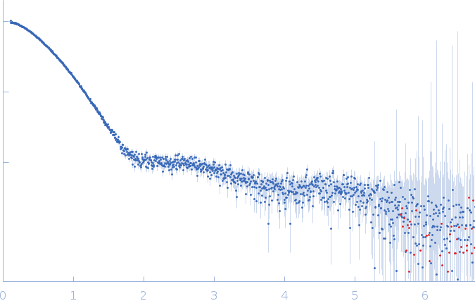
log I(s) 7.51×10⁴ 7.51×10³ 7.51×10² 7.51×101
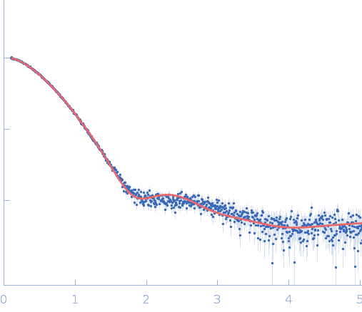
INTERLEUKIN 8 Heparin 8-mer small angle scattering data  s, nm-1
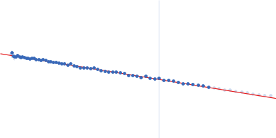
ln I(s)
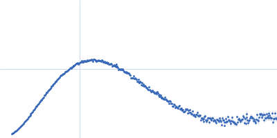
INTERLEUKIN 8 Heparin 8-mer Guinier plot ln 7.51×10⁴ Rg: 2.6 nm 0 (2.6 nm)-2 s2
(sRg)2I(s)/I(0)
INTERLEUKIN 8 Heparin 8-mer Kratky plot 1.104 0 3 sRg
p(r)
INTERLEUKIN 8 Heparin 8-mer pair distance distribution function Rg: 2.7 nm 0 Dmax: 11 nm

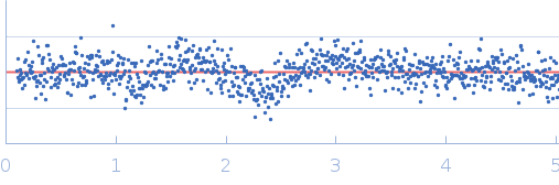
Data validation


Fits and models


log I(s)
 s, nm-1
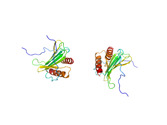
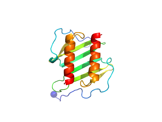
INTERLEUKIN 8 Heparin 8-mer CUSTOM IN-HOUSE model

SAXS data from solutions of Interleukin-8-Heparin sulfate (CXCL8:Heparin) in 20 mM sodium succinate, 100 mM NaCl, 5mM MgCl2, pH 5.5 were collected on the Rigaku BioSAXS-1000 instrument (Sealy Center For Structural Biology, UTMB-G, Galveston, TX, USA) using a Pilatus 100K detector at a sample-detector distance of 0.5 m and at a wavelength of λ = 0.15418 nm (I(s) vs s, where s = 4πsinθ/λ, and 2θ is the scattering angle). One solute concentration of 3.00 mg/ml was measured at 5°C. 10 successive 3600 second frames were collected. The data were normalized to the intensity of the transmitted beam and radially averaged; the scattering of the solvent-blank was subtracted.

INTERLEUKIN 8 (CXCL8)
Mol. type   Protein
Organism   Homo sapiens
Olig. state   Dimer
Mon. MW   8.0 kDa
 
UniProt   P10145 (28-99)
Sequence   FASTA
 
Heparin 8-mer (Hep08)
Mol. type   Other
Olig. state   Monomer
Mon. MW   2.4 kDa