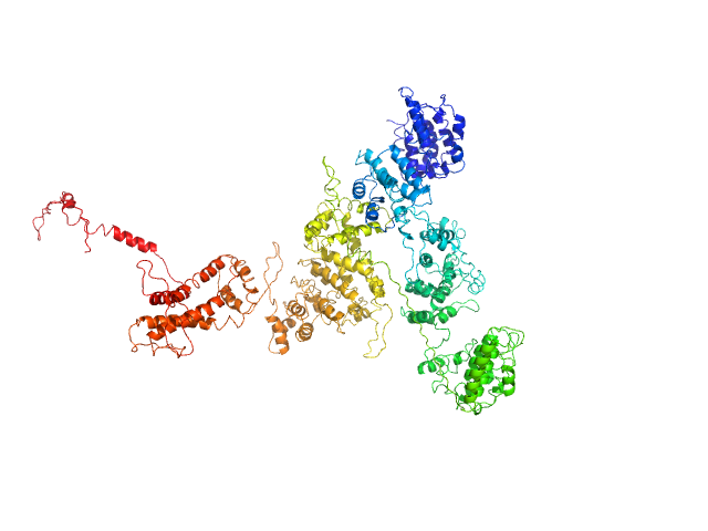
Search

 
Advanced search  

60 hits found for White

SASDH62 – Isoform 3 of Rap guanine nucleotide exchange factor 4, aE2F-DB08

Isoform 3 of Rap guanine nucleotide exchange factor 4 experimental SAS data
Isoform 3 of Rap guanine nucleotide exchange factor 4, aE2F-DB08 Rg histogram
Sample: Isoform 3 of Rap guanine nucleotide exchange factor 4 monomer, 114 kDa Mus musculus protein
Buffer: 150 mM NaCl, 1 mM EDTA, 1 mM DTT, and 10 mM Tris-HCl, pH: 7.5
Experiment: SAXS data collected at Anton Paar SAXSess, University of Utah on 2008 Aug 7
Mechanism of Epac activation: structural and functional analyses of Epac2 hinge mutants with constitutive and reduced activities. J Biol Chem 284(35):23644-51 (2009)
...White MA, Cheng X
RgGuinier 3.2 nm
Dmax 10.7 nm
VolumePorod 123 nm3

SASDH72 – Isoform 3 of Rap guanine nucleotide exchange factor 4, aE2G-DB08

Isoform 3 of Rap guanine nucleotide exchange factor 4 experimental SAS data
Isoform 3 of Rap guanine nucleotide exchange factor 4, aE2G-DB08 Rg histogram
Sample: Isoform 3 of Rap guanine nucleotide exchange factor 4 monomer, 114 kDa Mus musculus protein
Buffer: 150 mM NaCl, 1 mM EDTA, 1 mM DTT, and 10 mM Tris-HCl, pH: 7.5
Experiment: SAXS data collected at Anton Paar SAXSess, University of Utah on 2008 Aug 8
Mechanism of Epac activation: structural and functional analyses of Epac2 hinge mutants with constitutive and reduced activities. J Biol Chem 284(35):23644-51 (2009)
...White MA, Cheng X
RgGuinier 3.8 nm
Dmax 12.5 nm
VolumePorod 151 nm3

SASDH82 – Isoform 3 of Rap guanine nucleotide exchange factor 4, aE2W-DB08

Isoform 3 of Rap guanine nucleotide exchange factor 4 experimental SAS data
Isoform 3 of Rap guanine nucleotide exchange factor 4, aE2W-DB08 Rg histogram
Sample: Isoform 3 of Rap guanine nucleotide exchange factor 4 monomer, 114 kDa Mus musculus protein
Buffer: 150 mM NaCl, 1 mM EDTA, 1 mM DTT, and 10 mM Tris-HCl, pH: 7.5
Experiment: SAXS data collected at Anton Paar SAXSess, University of Utah on 2008 Aug 11
Mechanism of Epac activation: structural and functional analyses of Epac2 hinge mutants with constitutive and reduced activities. J Biol Chem 284(35):23644-51 (2009)
...White MA, Cheng X
RgGuinier 3.6 nm
Dmax 11.4 nm
VolumePorod 145 nm3

SASDLD2 – Circular permutant Carboxypeptidase G2-CP-N89-K177A

Carboxypeptidase G2 (circular permutant CP-N89) K177A experimental SAS data
PDB (PROTEIN DATA BANK) model
Sample: Carboxypeptidase G2 (circular permutant CP-N89) K177A dimer, 85 kDa Pseudomonas sp. (strain … protein
Buffer: 50 mM Tris, 100 mM NaCl, pH: 7.4
Experiment: SAXS data collected at 12.3.1 (SIBYLS), Advanced Light Source (ALS) on 2017 Jun 6
Massively parallel, computationally guided design of a proenzyme. Proc Natl Acad Sci U S A 119(15):e2116097119 (2022)
...White RE 3rd, Minetti CASA, Remeta DP, Tan VM, Drake JM, Khare SD
RgGuinier 3.6 nm
Dmax 14.0 nm
VolumePorod 105 nm3

SASDLE2 – Circular permutant Carboxypeptidase G2-CP-N89-K177A with methotrexate

Carboxypeptidase G2 (circular permutant CP-N89) K177AMethotrexate experimental SAS data
ROSETTA model
Sample: Carboxypeptidase G2 (circular permutant CP-N89) K177A dimer, 85 kDa Pseudomonas sp. (strain … protein
Methotrexate dimer, 1 kDa
Buffer: 50 mM Tris, 100 mM NaCl, pH: 7.4
Experiment: SAXS data collected at 12.3.1 (SIBYLS), Advanced Light Source (ALS) on 2017 Jun 6
Massively parallel, computationally guided design of a proenzyme. Proc Natl Acad Sci U S A 119(15):e2116097119 (2022)
...White RE 3rd, Minetti CASA, Remeta DP, Tan VM, Drake JM, Khare SD
RgGuinier 3.5 nm
Dmax 12.7 nm
VolumePorod 104 nm3

SASDLF2 – Pro-CPG2-1 (pro-enzyme design 1 of circular permutant Carboxypeptidase G2-CP-N89-K177A)

Pro-Carboxypeptidase G2 (circular permutant CP-N89) K177A Design 1 experimental SAS data
ROSETTA model
Sample: Pro-Carboxypeptidase G2 (circular permutant CP-N89) K177A Design 1 dimer, 100 kDa Pseudomonas sp. (strain … protein
Buffer: 50 mM Tris, 100 mM NaCl, pH: 7.4
Experiment: SAXS data collected at 12.3.1 (SIBYLS), Advanced Light Source (ALS) on 2018 May 8
Massively parallel, computationally guided design of a proenzyme. Proc Natl Acad Sci U S A 119(15):e2116097119 (2022)
...White RE 3rd, Minetti CASA, Remeta DP, Tan VM, Drake JM, Khare SD
RgGuinier 3.6 nm
Dmax 12.4 nm
VolumePorod 125 nm3

SASDLG2 – Pro-CPG2-1 (pro-enzyme design 1 of circular permutant Carboxypeptidase G2-CP-N89-K177A) with Methotrexate

MethotrexatePro-Carboxypeptidase G2 (circular permutant CP-N89) K177A Design 1 experimental SAS data
ROSETTA model
Sample: Methotrexate dimer, 1 kDa
Pro-Carboxypeptidase G2 (circular permutant CP-N89) K177A Design 1 dimer, 100 kDa Pseudomonas sp. (strain … protein
Buffer: 50 mM Tris, 100 mM NaCl, pH: 7.4
Experiment: SAXS data collected at 12.3.1 (SIBYLS), Advanced Light Source (ALS) on 2018 May 8
Massively parallel, computationally guided design of a proenzyme. Proc Natl Acad Sci U S A 119(15):e2116097119 (2022)
...White RE 3rd, Minetti CASA, Remeta DP, Tan VM, Drake JM, Khare SD
RgGuinier 3.5 nm
Dmax 12.2 nm
VolumePorod 123 nm3

SASDLH2 – Pro-CPG2-2 (pro-enzyme design 2 of circular permutant Carboxypeptidase G2-CP-N89-K177A)

Pro-Carboxypeptidase G2 (circular permutant CP-N89) K177A Design 2 experimental SAS data
ROSETTA model
Sample: Pro-Carboxypeptidase G2 (circular permutant CP-N89) K177A Design 2 dimer, 100 kDa Pseudomonas sp. (strain … protein
Buffer: 50 mM Tris, 100 mM NaCl, pH: 7.4
Experiment: SAXS data collected at 12.3.1 (SIBYLS), Advanced Light Source (ALS) on 2018 May 8
Massively parallel, computationally guided design of a proenzyme. Proc Natl Acad Sci U S A 119(15):e2116097119 (2022)
...White RE 3rd, Minetti CASA, Remeta DP, Tan VM, Drake JM, Khare SD
RgGuinier 3.7 nm
Dmax 12.5 nm
VolumePorod 126 nm3

SASDLJ2 – Pro-CPG2-2 (pro-enzyme design 2 of circular permutant Carboxypeptidase G2-CP-N89-K177A) with Methotrexate

MethotrexatePro-Carboxypeptidase G2 (circular permutant CP-N89) K177A Design 2 experimental SAS data
ROSETTA model
Sample: Methotrexate dimer, 1 kDa
Pro-Carboxypeptidase G2 (circular permutant CP-N89) K177A Design 2 dimer, 100 kDa Pseudomonas sp. (strain … protein
Buffer: 50 mM Tris, 100 mM NaCl, pH: 7.4
Experiment: SAXS data collected at 12.3.1 (SIBYLS), Advanced Light Source (ALS) on 2018 May 8
Massively parallel, computationally guided design of a proenzyme. Proc Natl Acad Sci U S A 119(15):e2116097119 (2022)
...White RE 3rd, Minetti CASA, Remeta DP, Tan VM, Drake JM, Khare SD
RgGuinier 3.5 nm
Dmax 12.1 nm
VolumePorod 125 nm3

SASDLK2 – Pro-CPG2-3 (pro-enzyme design 3 of circular permutant Carboxypeptidase G2-CP-N89-K177A)

Pro-Carboxypeptidase G2 (circular permutant CP-N89) K177A Design 3 experimental SAS data
ROSETTA model
Sample: Pro-Carboxypeptidase G2 (circular permutant CP-N89) K177A Design 3 dimer, 100 kDa Pseudomonas sp. (strain … protein
Buffer: 50 mM Tris, 100 mM NaCl, pH: 7.4
Experiment: SAXS data collected at 12.3.1 (SIBYLS), Advanced Light Source (ALS) on 2018 May 8
Massively parallel, computationally guided design of a proenzyme. Proc Natl Acad Sci U S A 119(15):e2116097119 (2022)
...White RE 3rd, Minetti CASA, Remeta DP, Tan VM, Drake JM, Khare SD
RgGuinier 3.7 nm
Dmax 13.0 nm
VolumePorod 127 nm3

SASDLL2 – Pro-CPG2-3 (pro-enzyme design 3 of circular permutant Carboxypeptidase G2-CP-N89-K177A) with Methotrexate

MethotrexatePro-Carboxypeptidase G2 (circular permutant CP-N89) K177A Design 3 experimental SAS data
ROSETTA model
Sample: Methotrexate dimer, 1 kDa
Pro-Carboxypeptidase G2 (circular permutant CP-N89) K177A Design 3 dimer, 100 kDa Pseudomonas sp. (strain … protein
Buffer: 50 mM Tris, 100 mM NaCl, pH: 7.4
Experiment: SAXS data collected at 12.3.1 (SIBYLS), Advanced Light Source (ALS) on 2018 May 8
Massively parallel, computationally guided design of a proenzyme. Proc Natl Acad Sci U S A 119(15):e2116097119 (2022)
...White RE 3rd, Minetti CASA, Remeta DP, Tan VM, Drake JM, Khare SD
RgGuinier 3.5 nm
Dmax 12.3 nm
VolumePorod 125 nm3

SASDLM2 – Pro-CPG2-1-Disulfide (pro-enzyme design 1 disulfide variant of circular permutant Carboxypeptidase G2-CP-N89-K177A)

Pro-Carboxypeptidase G2 (circular permutant CP-N89) K177A Design 1 Disulfide Variant experimental SAS data
ROSETTA model
Sample: Pro-Carboxypeptidase G2 (circular permutant CP-N89) K177A Design 1 Disulfide Variant dimer, 96 kDa Pseudomonas sp. (strain … protein
Buffer: 50 mM Tris, 100 mM NaCl, pH: 7.4
Experiment: SAXS data collected at 12.3.1 (SIBYLS), Advanced Light Source (ALS) on 2018 Dec 6
Massively parallel, computationally guided design of a proenzyme. Proc Natl Acad Sci U S A 119(15):e2116097119 (2022)
...White RE 3rd, Minetti CASA, Remeta DP, Tan VM, Drake JM, Khare SD
RgGuinier 3.6 nm
Dmax 12.6 nm
VolumePorod 120 nm3

SASDLN2 – Pro-CPG2-1-Disulfide (pro-enzyme design 1 disulfide variant of circular permutant Carboxypeptidase G2-CP-N89-K177A) with Methotrexate

MethotrexatePro-Carboxypeptidase G2 (circular permutant CP-N89) K177A Design 1 Disulfide Variant experimental SAS data
ROSETTA model
Sample: Methotrexate dimer, 1 kDa
Pro-Carboxypeptidase G2 (circular permutant CP-N89) K177A Design 1 Disulfide Variant dimer, 96 kDa Pseudomonas sp. (strain … protein
Buffer: 50 mM Tris, 100 mM NaCl, pH: 7.4
Experiment: SAXS data collected at 12.3.1 (SIBYLS), Advanced Light Source (ALS) on 2018 Dec 6
Massively parallel, computationally guided design of a proenzyme. Proc Natl Acad Sci U S A 119(15):e2116097119 (2022)
...White RE 3rd, Minetti CASA, Remeta DP, Tan VM, Drake JM, Khare SD
RgGuinier 3.5 nm
Dmax 12.3 nm
VolumePorod 125 nm3

SASDHW2 – The Legionella pneumophila type II secretion system substrate NttE

Uncharacterized protein experimental SAS data
GASBOR model
Sample: Uncharacterized protein dimer, 66 kDa Legionella pneumophila protein
Buffer: 20 mM Tris, 200 mM NaCl, pH: 8
Experiment: SAXS data collected at B21, Diamond Light Source on 2019 Sep 18
Structure, Dynamics and Cellular Insight Into Novel Substrates of the Legionella pneumophila Type II Secretion System Frontiers in Molecular Biosciences 7 (2020)
...White R, McIntire I, Sewell L, Richardson K, Shaw R, Pandini A, Cianciotto N, Garnett J
RgGuinier 2.9 nm
Dmax 9.4 nm
VolumePorod 114 nm3

SASDJH3 – ...White Lysozyme in presence of 100 mM 5-methyl uridine

Lysozyme C experimental SAS data
Lysozyme C Kratky plot
Sample: Lysozyme C monomer, 16 kDa Gallus gallus protein
Buffer: 40 mM sodium acetate, 150 mM NaCl, 100 mM 5-methyl uridine, pH: 3.8
Experiment: SAXS data collected at BL11 - NCD, ALBA on 2019 Jun 7
Improving data quality and expanding BioSAXS experiments to low-molecular-weight and low-concentration protein samples. Acta Crystallogr D Struct Biol 76(Pt 10):971-981 (2020)
Castellví A, Pascual-Izarra C, Crosas E, Malfois M, Juanhuix J
RgGuinier 1.5 nm
Dmax 4.2 nm
VolumePorod 21 nm3

SASDDP3 – N-propargyl glycine-Inactivated Proline utilization A from Bradyrhizobium diazoefficiens (formerly Bradyrhizobium japonicum) collected by SEC-SAXS

Bifunctional protein PutA experimental SAS data
PDB (PROTEIN DATA BANK) model
Sample: Bifunctional protein PutA dimer, 215 kDa Bradyrhizobium diazoefficiens protein
Buffer: 50 mM Tris, 50 mM NaCl, 0.5 mM TCEP, 5% (v/v) glycerol, pH: 7.8
Experiment: SAXS data collected at BioCAT 18ID, Advanced Photon Source (APS), Argonne National Laboratory on 2017 Jul 16
Redox Modulation of Oligomeric State in Proline Utilization A. Biophys J 114(12):2833-2843 (2018)
...White TA, Becker DF, Tanner JJ
RgGuinier 4.6 nm
Dmax 14.4 nm
VolumePorod 324 nm3

SASDDQ3 – Proline utilization A from Bradyrhizobium diazoefficiens (formerly Bradyrhizobium japonicum) collected by SEC-SAXS

Bifunctional protein PutA experimental SAS data
PDB (PROTEIN DATA BANK) model
Sample: Bifunctional protein PutA tetramer, 430 kDa Bradyrhizobium diazoefficiens protein
Buffer: 50 mM Tris, 50 mM NaCl, 0.5 mM TCEP, 5% (v/v) glycerol, pH: 7.8
Experiment: SAXS data collected at BioCAT 18ID, Advanced Photon Source (APS), Argonne National Laboratory on 2017 Jul 16
Redox Modulation of Oligomeric State in Proline Utilization A. Biophys J 114(12):2833-2843 (2018)
...White TA, Becker DF, Tanner JJ
RgGuinier 5.2 nm
Dmax 14.2 nm
VolumePorod 582 nm3

SASDG54 – Human macrophage mannose receptor 1 protein

Macrophage mannose receptor 1 experimental SAS data
ITASSER model
Sample: Macrophage mannose receptor 1 dimer, 315 kDa Mouse myeloma cell … protein
Buffer: 50mM Hepes, 100mM NaCl, 1mM DTT, pH: 7
Experiment: SAXS data collected at 12-ID-B SAXS/WAXS, Advanced Photon Source (APS), Argonne National Laboratory on 2016 Apr 15
Mannose receptor (CD206) activation in tumor-associated macrophages enhances adaptive and innate antitumor immune responses. Sci Transl Med 12(530) (2020)
...White J, Ravichandran S, Kumar P, Talsania K, Chen V, Ghebremedhin A, Karanam B, Bin Salam A, Amin R, Odzorig T, Aiken T, Nguyen V, Bian Y, Zarif JC, de Groot AE, Mehta M, Fan L, Hu X, Simeonov A, Pat...
RgGuinier 7.9 nm
Dmax 30.1 nm
VolumePorod 584 nm3

SASDLF4 – Human immunoglobulin gamma 2 (IgG2) ChiLob 7/4 F(ab)2 - Heavy chain C224S mutant

ChiLob 7/4 IgG2 F(ab)2 C224S mutant experimental SAS data
Human immunoglobulin gamma 2 (IgG2) ChiLob 7/4 F(ab)2 - Heavy chain C224S mutant Rg histogram
Sample: ChiLob 7/4 IgG2 F(ab)2 C224S mutant monomer, 99 kDa protein
Buffer: 50 mM HEPES, 150 mM KCl, pH: 7.5
Experiment: SAXS data collected at BM29, ESRF on 2018 Feb 9
Hinge disulfides in human IgG2 CD40 antibodies modulate receptor signaling by regulation of conformation and flexibility Science Immunology 7(73) (2022)
...White A, Essex J, Pearson A, Cragg M, Tews I
RgGuinier 3.9 nm
Dmax 14.2 nm
VolumePorod 135 nm3

SASDLG4 – Human immunoglobulin gamma 2 (IgG2) ChiLob 7/4 F(ab)2 - Heavy chain C224S+C225S mutant

ChiLob 7/4 IgG2 F(ab)2 C224S/C225S mutant experimental SAS data
Human immunoglobulin gamma 2 (IgG2) ChiLob 7/4 F(ab)2 - Heavy chain C224S+C225S mutant Rg histogram
Sample: ChiLob 7/4 IgG2 F(ab)2 C224S/C225S mutant monomer, 99 kDa protein
Buffer: 50 mM HEPES, 150 mM KCl, pH: 7.5
Experiment: SAXS data collected at BM29, ESRF on 2018 Feb 9
Hinge disulfides in human IgG2 CD40 antibodies modulate receptor signaling by regulation of conformation and flexibility Science Immunology 7(73) (2022)
...White A, Essex J, Pearson A, Cragg M, Tews I
RgGuinier 4.2 nm
Dmax 15.1 nm
VolumePorod 135 nm3

SASDLH4 – Human immunoglobulin gamma 2 (IgG2) ChiLob 7/4 F(ab)2 - Heavy chain C225S mutant

ChiLob 7/4 IgG2 F(ab)2 C225S mutant experimental SAS data
Human immunoglobulin gamma 2 (IgG2) ChiLob 7/4 F(ab)2 - Heavy chain C225S mutant Rg histogram
Sample: ChiLob 7/4 IgG2 F(ab)2 C225S mutant monomer, 99 kDa protein
Buffer: 50 mM HEPES, 150 mM KCl, pH: 7.5
Experiment: SAXS data collected at BM29, ESRF on 2018 Feb 9
Hinge disulfides in human IgG2 CD40 antibodies modulate receptor signaling by regulation of conformation and flexibility Science Immunology 7(73) (2022)
...White A, Essex J, Pearson A, Cragg M, Tews I
RgGuinier 4.0 nm
Dmax 14.5 nm
VolumePorod 131 nm3

SASDHJ4 – Dengue Virus 2 New Guinea C (DENV-2 C, capsid protein), apo form

Dengue Virus 2 New Guinea C experimental SAS data
CORAL model
Sample: Dengue Virus 2 New Guinea C dimer, 19 kDa Dengue virus 2 protein
Buffer: 100 mM NaCl, 25 mM HEPES, pH: 7.4
Experiment: SAXS data collected at Rigaku BioSAXS-1000, Sealy Center For Structural Biology, UTMB-G on 2018 Jul 5
A cocrystal structure of dengue capsid protein in complex of inhibitor. Proc Natl Acad Sci U S A 117(30):17992-18001 (2020)
...White MA, Shi PY
RgGuinier 1.8 nm
Dmax 5.4 nm
VolumePorod 52 nm3

SASDLJ4 – Human immunoglobulin gamma 2 (IgG2) ChiLob 7/4 F(ab)2 - Heavy chain C228S mutant

ChiLob 7/4 IgG2 F(ab)2 C228S mutant experimental SAS data
Human immunoglobulin gamma 2 (IgG2) ChiLob 7/4 F(ab)2 - Heavy chain C228S mutant Rg histogram
Sample: ChiLob 7/4 IgG2 F(ab)2 C228S mutant monomer, 99 kDa protein
Buffer: 50 mM HEPES, 150 mM KCl, pH: 7.5
Experiment: SAXS data collected at BM29, ESRF on 2018 Feb 9
Hinge disulfides in human IgG2 CD40 antibodies modulate receptor signaling by regulation of conformation and flexibility Science Immunology 7(73) (2022)
...White A, Essex J, Pearson A, Cragg M, Tews I
RgGuinier 4.1 nm
Dmax 13.5 nm
VolumePorod 135 nm3

SASDHK4 – Dengue Virus 2 New Guinea C (DENV-2 C, capsid protein) with saturating ST148 inhibitor

Dengue Virus 2 New Guinea C experimental SAS data
CORAL model
Sample: Dengue Virus 2 New Guinea C dimer, 19 kDa Dengue virus 2 protein
Buffer: 100 mM NaCl, 25 mM HEPES, pH 7.4, 10 µM ST148, pH: 7.4
Experiment: SAXS data collected at Rigaku BioSAXS-1000, Sealy Center For Structural Biology, UTMB-G on 2018 Jul 26
A cocrystal structure of dengue capsid protein in complex of inhibitor. Proc Natl Acad Sci U S A 117(30):17992-18001 (2020)
...White MA, Shi PY
RgGuinier 1.9 nm
Dmax 6.0 nm
VolumePorod 52 nm3

SASDLK4 – Human immunoglobulin gamma 2 (IgG2) ChiLob 7/4 F(ab)2 - Heavy chain C224S, kappa chain C214S mutant

ChiLob 7/4 IgG2 F(ab)2 C224S/kC214S mutant experimental SAS data
Human immunoglobulin gamma 2 (IgG2) ChiLob 7/4 F(ab)2 - Heavy chain C224S, kappa chain C214S mutant Rg histogram
Sample: ChiLob 7/4 IgG2 F(ab)2 C224S/kC214S mutant monomer, 99 kDa protein
Buffer: 50 mM HEPES, 150 mM KCl, pH: 7.5
Experiment: SAXS data collected at BM29, ESRF on 2018 Feb 9
Hinge disulfides in human IgG2 CD40 antibodies modulate receptor signaling by regulation of conformation and flexibility Science Immunology 7(73) (2022)
...White A, Essex J, Pearson A, Cragg M, Tews I
RgGuinier 4.0 nm
Dmax 13.3 nm
VolumePorod 133 nm3

SASDLL4 – Human immunoglobulin gamma 2 (IgG2) ChiLob 7/4 F(ab)2 - Heavy chain C225S, kappa chain C214S mutant

ChiLob 7/4 IgG2 F(ab)2 C225S/kC214S mutant experimental SAS data
Human immunoglobulin gamma 2 (IgG2) ChiLob 7/4 F(ab)2 - Heavy chain C225S, kappa chain C214S mutant Rg histogram
Sample: ChiLob 7/4 IgG2 F(ab)2 C225S/kC214S mutant monomer, 99 kDa protein
Buffer: 50 mM HEPES, 150 mM KCl, pH: 7.5
Experiment: SAXS data collected at BM29, ESRF on 2018 Feb 9
Hinge disulfides in human IgG2 CD40 antibodies modulate receptor signaling by regulation of conformation and flexibility Science Immunology 7(73) (2022)
...White A, Essex J, Pearson A, Cragg M, Tews I
RgGuinier 4.0 nm
Dmax 13.7 nm
VolumePorod 130 nm3

SASDQM4 – CRISPR associated Lon protease (CalpL)

SAVED domain-containing protein experimental SAS data
DAMMIF model
Sample: SAVED domain-containing protein monomer, 58 kDa Sulfurihydrogenibium sp. (strain … protein
Buffer: 20 mM Tris, 50 mM NaCl, pH: 8
Experiment: SAXS data collected at EMBL P12, PETRA III on 2021 Dec 14
Antiviral signalling by a cyclic nucleotide activated CRISPR protease. Nature 614(7946):168-174 (2023)
...White MF, Hagelueken G
RgGuinier 3.1 nm
Dmax 10.3 nm
VolumePorod 80 nm3

SASDQN4 – Complex of CRISPR associated Lon protease (CalpL) with a 10 kDa C-terminal fragment of CalpT a putative MazF-like toxin (CalpT10)

SAVED domain-containing proteinUncharacterized protein (putative MazF-like toxin) experimental SAS data
CUSTOM IN-HOUSE model
Sample: SAVED domain-containing protein monomer, 58 kDa Sulfurihydrogenibium sp. (strain … protein
Uncharacterized protein (putative MazF-like toxin) monomer, 9 kDa Sulfurihydrogenibium sp. (strain … protein
Buffer: 20 mM Tris, 50 mM NaCl, pH: 8
Experiment: SAXS data collected at EMBL P12, PETRA III on 2021 Dec 14
Antiviral signalling by a cyclic nucleotide activated CRISPR protease. Nature 614(7946):168-174 (2023)
...White MF, Hagelueken G
RgGuinier 3.4 nm
Dmax 11.7 nm
VolumePorod 93 nm3

SASDQP4 – CRISPR associated Lon protease (CalpL) in the presence of cyclic oligoadenylate cA4

SAVED domain-containing protein experimental SAS data
DAMMIF model
Sample: SAVED domain-containing protein monomer, 58 kDa Sulfurihydrogenibium sp. (strain … protein
Buffer: 20 mM Tris, 50 mM NaCl, pH: 8
Experiment: SAXS data collected at EMBL P12, PETRA III on 2022 Apr 8
Antiviral signalling by a cyclic nucleotide activated CRISPR protease. Nature 614(7946):168-174 (2023)
...White MF, Hagelueken G
RgGuinier 3.2 nm
Dmax 10.3 nm
VolumePorod 82 nm3

SASDQQ4 – CRISPR associated Lon protease (CalpL) asymmetrically dimerizing in the presence of cyclic oligoadenylate cA4

SAVED domain-containing protein experimental SAS data
SASREF MX model
Sample: SAVED domain-containing protein monomer, 58 kDa Sulfurihydrogenibium sp. (strain … protein
Buffer: 20 mM Tris, 50 mM NaCl, pH: 8
Experiment: SAXS data collected at EMBL P12, PETRA III on 2022 Apr 8
Antiviral signalling by a cyclic nucleotide activated CRISPR protease. Nature 614(7946):168-174 (2023)
...White MF, Hagelueken G
RgGuinier 3.8 nm
Dmax 13.0 nm
VolumePorod 116 nm3

SASDD95 – Neurexin 1a L5L6

Neurexin 1a L5L6 experimental SAS data
Neurexin 1a L5L6 Rg histogram
Sample: Neurexin 1a L5L6 monomer, 44 kDa protein
Buffer: 20 mM HEPES pH 8, 150 mM NaCl, 0.5mM CaCl2, pH: 8
Experiment: SAXS data collected at Rigaku BioSAXS-1000, Sealy Center For Structural Biology, UTMB-G on 2016 Sep 6
Structural Plasticity of Neurexin 1α: Implications for its Role as Synaptic Organizer. J Mol Biol 430(21):4325-4343 (2018)
...White MA, Ren G, Rudenko G
RgGuinier 3.0 nm
Dmax 10.0 nm
VolumePorod 69 nm3

SASDDA5 – Neurexin 1a L5L6 with ss6 insert

Neurexin 1a L5L6 with ss6 insert experimental SAS data
Neurexin 1a L5L6 with ss6 insert Rg histogram
Sample: Neurexin 1a L5L6 with ss6 insert monomer, 45 kDa Homo sapiens protein
Buffer: 20 mM HEPES pH 8, 150 mM NaCl, 0.5mM CaCl2, pH: 8
Experiment: SAXS data collected at Rigaku BioSAXS-1000, Sealy Center For Structural Biology, UTMB-G on 2016 Sep 26
Structural Plasticity of Neurexin 1α: Implications for its Role as Synaptic Organizer. J Mol Biol 430(21):4325-4343 (2018)
...White MA, Ren G, Rudenko G
RgGuinier 3.2 nm
Dmax 12.4 nm
VolumePorod 70 nm3

SASDMA5 – cone-rod homeobox protein (a peptide of the DNA binding domain of CRX)

Cone-rod homeobox protein experimental SAS data
OTHER model
Sample: Cone-rod homeobox protein monomer, 7 kDa Homo sapiens protein
Buffer: 50 mM sodium phosphate,100 mM NaCl, and 5 mM imidazole, pH: 7
Experiment: SAXS data collected at 12.3.1 (SIBYLS), Advanced Light Source (ALS) on 2021 Jun 15
Structural and functional analysis of the human Cone-rod homeobox transcription factor. Proteins (2022)
...White AM, Enke RA, Berndsen CE
RgGuinier 1.2 nm
Dmax 4.0 nm
VolumePorod 10 nm3

SASDJB6 – Lactococcus phage Phi28 apo-gp11 encapsidation protein

gp11 encapsidation protein experimental SAS data
OTHER model
Sample: gp11 encapsidation protein decamer, 446 kDa Lactococcus phage Phi28 protein
Buffer: 100 mM NaCl, 25 mM HEPES, pH: 7.4
Experiment: SAXS data collected at Rigaku BioSAXS-1000, Sealy Center For Structural Biology, UTMB-G on 2015 May 6
Lactococcus Phage ASCC Phi28 gp11
Mark White
RgGuinier 5.3 nm
Dmax 16.3 nm

SASDCQ6 – Rap guanine nucleotide exchange factor 3 (isoform3) - apo form

Rap guanine nucleotide exchange factor 3 experimental SAS data
SWISSMODEL model
Sample: Rap guanine nucleotide exchange factor 3 monomer, 100 kDa Homo sapiens protein
Buffer: 1mM EDTA, 10mM DTT, 500mM NaCl, and 10mM Tris, pH: 9
Experiment: SAXS data collected at Rigaku BioSAXS-1000, Sealy Center For Structural Biology, UTMB-G on 2012 Sep 7
Conformational States of Exchange Protein Directly Activated by cAMP (EPAC1) Revealed by Ensemble Modeling and Integrative Structural Biology. Cells 9(1) (2019)
White MA, Tsalkova T, Mei FC, Cheng X
RgGuinier 3.4 nm
Dmax 11.0 nm
VolumePorod 180 nm3

SASDCR6 – Rap guanine nucleotide exchange factor 3 (isoform3) - binary form with cAMP

Rap guanine nucleotide exchange factor 3 (dimer) experimental SAS data
CORAL model
Sample: Rap guanine nucleotide exchange factor 3 (dimer) dimer, 200 kDa Homo sapiens protein
Buffer: 1mM EDTA, 10mM DTT, 500mM NaCl, 1mM cAMP, and 10mM Tris, pH: 9
Experiment: SAXS data collected at Rigaku BioSAXS-1000, Sealy Center For Structural Biology, UTMB-G on 2012 Jan 30
Conformational States of Exchange Protein Directly Activated by cAMP (EPAC1) Revealed by Ensemble Modeling and Integrative Structural Biology. Cells 9(1) (2019)
White MA, Tsalkova T, Mei FC, Cheng X
RgGuinier 5.3 nm
Dmax 15.7 nm
VolumePorod 415 nm3

SASDCS6 – Rap guanine nucleotide exchange factor 3 (isoform3) bound to RAS related protein 1b - with cAMP

Rap guanine nucleotide exchange factor 3RAS related protein 1b experimental SAS data
SWISSMODEL model
Sample: Rap guanine nucleotide exchange factor 3 monomer, 100 kDa Homo sapiens protein
RAS related protein 1b monomer, 18 kDa Mus musculus protein
Buffer: 1mM EDTA, 10mM DTT, 500mM NaCl, 1mM cAMP, and 10mM Tris, pH: 9
Experiment: SAXS data collected at Rigaku BioSAXS-1000, Sealy Center For Structural Biology, UTMB-G on 2013 Apr 1
Conformational States of Exchange Protein Directly Activated by cAMP (EPAC1) Revealed by Ensemble Modeling and Integrative Structural Biology. Cells 9(1) (2019)
White MA, Tsalkova T, Mei FC, Cheng X
RgGuinier 4.1 nm
Dmax 14.2 nm
VolumePorod 207 nm3

SASDHN8 – Human tumor necrosis factor receptor superfamily member 5 (CD40) extracellular domain in complex with human immunoglobulin gamma 1 (IgG1) 341G2 F(ab)

Tumor necrosis factor receptor superfamily member 5Human 341G2 F(ab) experimental SAS data
HADDOCK model
Sample: Tumor necrosis factor receptor superfamily member 5 monomer, 19 kDa Homo sapiens protein
Human 341G2 F(ab) monomer, 73 kDa Homo sapiens protein
Buffer: Phosphate-buffered saline, pH: 7
Experiment: SAXS data collected at B21, Diamond Light Source on 2019 Sep 18
Isotype Switching Converts Anti-CD40 Antagonism to Agonism to Elicit Potent Antitumor Activity Cancer Cell (2020)
...White A, Tews I, Glennie M, Cragg M
RgGuinier 3.7 nm
Dmax 13.4 nm
VolumePorod 97 nm3

SASDDV8 – Boiled chicken egg albumen

Ovalbumin experimental SAS data
Ovalbumin Kratky plot
Sample: Ovalbumin monomer, 43 kDa Gallus gallus protein
Buffer: Water, pH: 7
Experiment: SAXS data collected at Bruker Nonius FR591, University of Pennslyvania on 2013 Jun 27
The Proof Is in the Pidan: Generalizing Proteins as Patchy Particles. ACS Cent Sci 4(7):840-853 (2018)
Cai J, Sweeney AM

SASDDW8 – Chinese century egg albumen (pidan) made from quail egg

Ovalbumin (common quail) experimental SAS data
Ovalbumin (common quail) Kratky plot
Sample: Ovalbumin (common quail) monomer, 42 kDa Coturnix coturnix protein
Buffer: Water, pH: 7
Experiment: SAXS data collected at Bruker Nonius FR591, University of Pennslyvania on 2013 Jun 27
The Proof Is in the Pidan: Generalizing Proteins as Patchy Particles. ACS Cent Sci 4(7):840-853 (2018)
Cai J, Sweeney AM

SASDDX8 – Raw chicken egg albumen

Ovalbumin experimental SAS data
Ovalbumin Kratky plot
Sample: Ovalbumin monomer, 43 kDa Gallus gallus protein
Buffer: Water, pH: 7
Experiment: SAXS data collected at Bruker Nonius FR591, University of Pennslyvania on 2013 Jun 27
The Proof Is in the Pidan: Generalizing Proteins as Patchy Particles. ACS Cent Sci 4(7):840-853 (2018)
Cai J, Sweeney AM

SASDDY8 – Chicken ovalbumin gel at high pH

Ovalbumin experimental SAS data
DAMMIF model
Sample: Ovalbumin monomer, 43 kDa Gallus gallus protein
Buffer: 0.16 M NaOH, pH: 13.2
Experiment: SAXS data collected at X9A, National Synchrotron Light Source (NSLS) on 2014 Feb 21
The Proof Is in the Pidan: Generalizing Proteins as Patchy Particles. ACS Cent Sci 4(7):840-853 (2018)
Cai J, Sweeney AM

SASDD29 – Low load concentration of apo alpha-aminoadipic semialdehyde dehydrogenase (ALDH7A1) collected by SEC-SAXS

Alpha-aminoadipic semialdehyde dehydrogenase experimental SAS data
PDB (PROTEIN DATA BANK) model
Sample: Alpha-aminoadipic semialdehyde dehydrogenase tetramer, 222 kDa Homo sapiens protein
Buffer: 50 mM Tris, 50 mM NaCl, 0.5 mM DTT, 5% (v/v) glycerol, pH: 7.8
Experiment: SAXS data collected at BioCAT 18ID, Advanced Photon Source (APS), Argonne National Laboratory on 2018 Feb 22
NAD+ Promotes Assembly of the Active Tetramer of Aldehyde Dehydrogenase 7A1. FEBS Lett (2018)
...White TA, Chakravarthy S, Tanner JJ
RgGuinier 3.5 nm
VolumePorod 212 nm3

SASDD39 – Medium load concentration of apo alpha-aminoadipic semialdehyde dehydrogenase (ALDH7A1) collected by SEC-SAXS

Alpha-aminoadipic semialdehyde dehydrogenase experimental SAS data
PDB (PROTEIN DATA BANK) model
Sample: Alpha-aminoadipic semialdehyde dehydrogenase tetramer, 222 kDa Homo sapiens protein
Buffer: 50 mM Tris, 50 mM NaCl, 0.5 mM DTT, 5% (v/v) glycerol, pH: 7.8
Experiment: SAXS data collected at BioCAT 18ID, Advanced Photon Source (APS), Argonne National Laboratory on 2018 Feb 22
NAD+ Promotes Assembly of the Active Tetramer of Aldehyde Dehydrogenase 7A1. FEBS Lett (2018)
...White TA, Chakravarthy S, Tanner JJ
RgGuinier 3.7 nm
VolumePorod 229 nm3

SASDD49 – High load concentration of apo alpha-aminoadipic semialdehyde dehydrogenase (ALDH7A1) collected by SEC-SAXS

Alpha-aminoadipic semialdehyde dehydrogenase experimental SAS data
PDB (PROTEIN DATA BANK) model
Sample: Alpha-aminoadipic semialdehyde dehydrogenase tetramer, 222 kDa Homo sapiens protein
Buffer: 50 mM Tris, 50 mM NaCl, 0.5 mM DTT, 5% (v/v) glycerol, pH: 7.8
Experiment: SAXS data collected at BioCAT 18ID, Advanced Photon Source (APS), Argonne National Laboratory on 2018 Feb 22
NAD+ Promotes Assembly of the Active Tetramer of Aldehyde Dehydrogenase 7A1. FEBS Lett (2018)
...White TA, Chakravarthy S, Tanner JJ
RgGuinier 3.7 nm
VolumePorod 238 nm3

SASDD59 – Low load concentration of alpha-aminoadipic semialdehyde dehydrogenase (ALDH7A1) with nicotinamide adenine dinucleotide (NAD) collected by SEC-SAXS

Alpha-aminoadipic semialdehyde dehydrogenase experimental SAS data
PDB (PROTEIN DATA BANK) model
Sample: Alpha-aminoadipic semialdehyde dehydrogenase tetramer, 222 kDa Homo sapiens protein
Buffer: 50 mM Tris, 50 mM NaCl, 0.5 mM DTT, 5% (v/v) glycerol, 1 mM NAD, pH: 7.8
Experiment: SAXS data collected at BioCAT 18ID, Advanced Photon Source (APS), Argonne National Laboratory on 2018 Feb 22
NAD+ Promotes Assembly of the Active Tetramer of Aldehyde Dehydrogenase 7A1. FEBS Lett (2018)
...White TA, Chakravarthy S, Tanner JJ
RgGuinier 3.7 nm
VolumePorod 277 nm3

SASDD69 – Medium load concentration of alpha-aminoadipic semialdehyde dehydrogenase (ALDH7A1) with nicotinamide adenine dinucleotide (NAD) collected by SEC-SAXS

Alpha-aminoadipic semialdehyde dehydrogenase experimental SAS data
PDB (PROTEIN DATA BANK) model
Sample: Alpha-aminoadipic semialdehyde dehydrogenase tetramer, 222 kDa Homo sapiens protein
Buffer: 50 mM Tris, 50 mM NaCl, 0.5 mM DTT, 5% (v/v) glycerol, 1 mM NAD, pH: 7.8
Experiment: SAXS data collected at BioCAT 18ID, Advanced Photon Source (APS), Argonne National Laboratory on 2018 Feb 22
NAD+ Promotes Assembly of the Active Tetramer of Aldehyde Dehydrogenase 7A1. FEBS Lett (2018)
...White TA, Chakravarthy S, Tanner JJ
RgGuinier 3.8 nm
VolumePorod 275 nm3

SASDD79 – High load concentration of alpha-aminoadipic semialdehyde dehydrogenase ALDH7A1 with nicotinamide adenine dinucleotide (NAD) collected by SEC-SAXS

Alpha-aminoadipic semialdehyde dehydrogenase experimental SAS data
PDB (PROTEIN DATA BANK) model
Sample: Alpha-aminoadipic semialdehyde dehydrogenase tetramer, 222 kDa Homo sapiens protein
Buffer: 50 mM Tris, 50 mM NaCl, 0.5 mM DTT, 5% (v/v) glycerol, 1 mM NAD, pH: 7.8
Experiment: SAXS data collected at BioCAT 18ID, Advanced Photon Source (APS), Argonne National Laboratory on 2018 Feb 22
NAD+ Promotes Assembly of the Active Tetramer of Aldehyde Dehydrogenase 7A1. FEBS Lett (2018)
...White TA, Chakravarthy S, Tanner JJ
RgGuinier 3.8 nm
VolumePorod 277 nm3

SASDJB9 – LIM/homeobox protein Lhx3 homeodomain (Lhx3-HD): HD3

LIM/homeobox protein Lhx3 experimental SAS data
MONSA model
Sample: LIM/homeobox protein Lhx3 monomer, 10 kDa Mus musculus protein
Buffer: 20 mM sodium phosphate monobasic/dibasic, 100 mM NaCl, 1 mM DTT, pH: 7.4
Experiment: SAXS data collected at SAXS/WAXS, Australian Synchrotron on 2018 Nov 2
Contrasting DNA-binding behaviour by ISL1 and LHX3 underpins differential gene targeting in neuronal cell specification Journal of Structural Biology: X :100043 (2020)
...White L, Kwan A, Trewhella J, Matthews J
RgGuinier 2.1 nm
Dmax 7.0 nm
VolumePorod 18 nm3

SASDJC9 – Insulin gene enhancer protein Isl-1 LIM-interaction domain (Isl1-LID) fused to the LIM/homeobox protein Lhx3 LIM-homeodomain (Lhx3-LIMHD): LLHD3

LIM/homeobox protein Lhx3Insulin gene enhancer protein ISL-1 experimental SAS data
MONSA model
Sample: LIM/homeobox protein Lhx3 monomer, 23 kDa Mus musculus protein
Insulin gene enhancer protein ISL-1 monomer, 4 kDa Mus musculus protein
Buffer: 20 mM sodium phosphate monobasic/dibasic, 100 mM NaCl, 1 mM DTT, pH: 7.4
Experiment: SAXS data collected at SAXS/WAXS, Australian Synchrotron on 2018 Nov 2
Contrasting DNA-binding behaviour by ISL1 and LHX3 underpins differential gene targeting in neuronal cell specification Journal of Structural Biology: X :100043 (2020)
...White L, Kwan A, Trewhella J, Matthews J
RgGuinier 3.3 nm
Dmax 12.0 nm
VolumePorod 39 nm3

SASDJD9 – Insulin gene enhancer protein Isl-1 homeodomain (Isl1-HD) fused to the LIM/homeobox protein Lhx3 homeodomain (Lhx3-HD): 2HD

Insulin gene enhancer protein ISL-1LIM/homeobox protein Lhx3 experimental SAS data
MONSA model
Sample: Insulin gene enhancer protein ISL-1 monomer, 12 kDa Mus musculus protein
LIM/homeobox protein Lhx3 monomer, 9 kDa Mus musculus protein
Buffer: 20 mM sodium phosphate monobasic/dibasic, 100 mM NaCl, 1 mM DTT, pH: 7.4
Experiment: SAXS data collected at SAXS/WAXS, Australian Synchrotron on 2018 Nov 2
Contrasting DNA-binding behaviour by ISL1 and LHX3 underpins differential gene targeting in neuronal cell specification Journal of Structural Biology: X :100043 (2020)
...White L, Kwan A, Trewhella J, Matthews J
RgGuinier 3.4 nm
Dmax 12.7 nm
VolumePorod 40 nm3

SASDJE9 – Insulin gene enhancer protein Isl-1 homeodomain/LIM-interaction domain (Isl1-HDLID) fused to the LIM/homeobox protein Lhx3 LIM-homeodomain (Lhx3-LIMHD): 2HDLL

Insulin gene enhancer protein ISL-1LIM/homeobox protein Lhx3 experimental SAS data
MONSA model
Sample: Insulin gene enhancer protein ISL-1 monomer, 14 kDa Mus musculus protein
LIM/homeobox protein Lhx3 monomer, 23 kDa Mus musculus protein
Buffer: 20 mM sodium phosphate monobasic/dibasic, 100 mM NaCl, 1 mM DTT, pH: 7.4
Experiment: SAXS data collected at SAXS/WAXS, Australian Synchrotron on 2018 Nov 2
Contrasting DNA-binding behaviour by ISL1 and LHX3 underpins differential gene targeting in neuronal cell specification Journal of Structural Biology: X :100043 (2020)
...White L, Kwan A, Trewhella J, Matthews J
RgGuinier 3.4 nm
Dmax 13.0 nm
VolumePorod 76 nm3

SASDJF9 – M100 DNA oligonucleotide bound to the LIM/homeobox protein Lhx3 homeodomain (Lhx3-HD): M100 + HD3

M100 oligonucleotideLIM/homeobox protein Lhx3 experimental SAS data
MONSA model
Sample: M100 oligonucleotide monomer, 12 kDa DNA
LIM/homeobox protein Lhx3 monomer, 10 kDa Mus musculus protein
Buffer: 20 mM sodium phosphate monobasic/dibasic, 100 mM NaCl, 1 mM DTT, pH: 7.4
Experiment: SAXS data collected at SAXS/WAXS, Australian Synchrotron on 2018 Nov 2
Contrasting DNA-binding behaviour by ISL1 and LHX3 underpins differential gene targeting in neuronal cell specification Journal of Structural Biology: X :100043 (2020)
...White L, Kwan A, Trewhella J, Matthews J
RgGuinier 2.1 nm
Dmax 7.0 nm
VolumePorod 27 nm3

SASDJG9 – M100 DNA oligonucleotide bound to the protein fusion of Insulin gene enhancer protein Isl-1 LIM-interaction domain (Isl1-LID) and LIM/homeobox protein Lhx3 LIM-homeodomain (Lhx3-LIMHD): M100 + LLHD3

M100 oligonucleotideLIM/homeobox protein Lhx3Insulin gene enhancer protein ISL-1 experimental SAS data
MONSA model
Sample: M100 oligonucleotide monomer, 12 kDa DNA
LIM/homeobox protein Lhx3 monomer, 23 kDa Mus musculus protein
Insulin gene enhancer protein ISL-1 monomer, 4 kDa Mus musculus protein
Buffer: 20 mM sodium phosphate monobasic/dibasic, 100 mM NaCl, 1 mM DTT, pH: 7.4
Experiment: SAXS data collected at SAXS/WAXS, Australian Synchrotron on 2018 Nov 2
Contrasting DNA-binding behaviour by ISL1 and LHX3 underpins differential gene targeting in neuronal cell specification Journal of Structural Biology: X :100043 (2020)
...White L, Kwan A, Trewhella J, Matthews J
RgGuinier 3.6 nm
Dmax 13.0 nm
VolumePorod 49 nm3

SASDJH9 – M100 DNA oligonucleotide bound to the protein fusion of Insulin gene enhancer protein Isl-1 homeodomain (Isl1-HD) and LIM/homeobox protein Lhx3 homeodomain (Lhx3-HD): M100 + 2HD

M100 oligonucleotideInsulin gene enhancer protein ISL-1LIM/homeobox protein Lhx3 experimental SAS data
MONSA model
Sample: M100 oligonucleotide monomer, 12 kDa DNA
Insulin gene enhancer protein ISL-1 monomer, 12 kDa Mus musculus protein
LIM/homeobox protein Lhx3 monomer, 9 kDa Mus musculus protein
Buffer: 20 mM sodium phosphate monobasic/dibasic, 100 mM NaCl, 1 mM DTT, pH: 7.4
Experiment: SAXS data collected at SAXS/WAXS, Australian Synchrotron on 2018 Nov 2
Contrasting DNA-binding behaviour by ISL1 and LHX3 underpins differential gene targeting in neuronal cell specification Journal of Structural Biology: X :100043 (2020)
...White L, Kwan A, Trewhella J, Matthews J
RgGuinier 2.4 nm
Dmax 7.5 nm
VolumePorod 41 nm3

SASDJJ9 – M100 DNA oligonucleotide bound to the protein fusion of Insulin gene enhancer protein Isl-1 homeodomain/LIM-interaction domain (Isl1-HDLID) and LIM/homeobox protein Lhx3 LIM-homeodomain (Lhx3-LIMHD): ...

M100 oligonucleotideInsulin gene enhancer protein ISL-1LIM/homeobox protein Lhx3 experimental SAS data
MONSA model
Sample: M100 oligonucleotide monomer, 12 kDa DNA
Insulin gene enhancer protein ISL-1 monomer, 14 kDa Mus musculus protein
LIM/homeobox protein Lhx3 monomer, 23 kDa Mus musculus protein
Buffer: 20 mM sodium phosphate monobasic/dibasic, 100 mM NaCl, 1 mM DTT, pH: 7.4
Experiment: SAXS data collected at SAXS/WAXS, Australian Synchrotron on 2018 Nov 2
Contrasting DNA-binding behaviour by ISL1 and LHX3 underpins differential gene targeting in neuronal cell specification Journal of Structural Biology: X :100043 (2020)
...White L, Kwan A, Trewhella J, Matthews J
RgGuinier 3.6 nm
Dmax 14.0 nm
VolumePorod 70 nm3

SASDJK9 – M100 DNA oligonucleotide

M100 oligonucleotide experimental SAS data
MONSA model
Sample: M100 oligonucleotide monomer, 12 kDa DNA
Buffer: 20 mM sodium phosphate monobasic/dibasic, 100 mM NaCl, 1 mM DTT, pH: 7.4
Experiment: SAXS data collected at SAXS/WAXS, Australian Synchrotron on 2018 Nov 2
Contrasting DNA-binding behaviour by ISL1 and LHX3 underpins differential gene targeting in neuronal cell specification Journal of Structural Biology: X :100043 (2020)
...White L, Kwan A, Trewhella J, Matthews J
RgGuinier 1.9 nm
Dmax 6.8 nm
VolumePorod 15 nm3

SASDDY9 – Protein translocase subunit SecA (full length, amino acids 1-901)

Protein translocase subunit SecA experimental SAS data
Protein translocase subunit SecA Kratky plot
Sample: Protein translocase subunit SecA dimer, 204 kDa Escherichia coli protein
Buffer: 20mM HEPES, 100mM NaCl, 1mM TCEP, pH: 8
Experiment: SAXS data collected at BM29, ESRF on 2016 Jul 18
The C-terminal tail of the bacterial translocation ATPase SecA modulates its activity. Elife 8 (2019)
...White SA, Ward DG, Mohammed F, Rahman KF, Wynne M, Hughes GW, Kramer G, Bukau B, Huber D
RgGuinier 4.2 nm
Dmax 14.9 nm
VolumePorod 424 nm3

SASDDZ9 – Protein translocase subunit SecA (amino acids 1-880)

Protein translocase subunit SecA experimental SAS data
Protein translocase subunit SecA Kratky plot
Sample: Protein translocase subunit SecA dimer, 199 kDa Escherichia coli protein
Buffer: 20mM HEPES, 100mM NaCl, 1mM TCEP, pH: 8
Experiment: SAXS data collected at BM29, ESRF on 2016 Jul 18
The C-terminal tail of the bacterial translocation ATPase SecA modulates its activity. Elife 8 (2019)
...White SA, Ward DG, Mohammed F, Rahman KF, Wynne M, Hughes GW, Kramer G, Bukau B, Huber D
RgGuinier 4.2 nm
Dmax 14.8 nm
VolumePorod 380 nm3

SASDE22 – Protein translocase subunit SecA (amino acids 1-832)

Protein translocase subunit SecA experimental SAS data
Protein translocase subunit SecA Kratky plot
Sample: Protein translocase subunit SecA dimer, 189 kDa Escherichia coli protein
Buffer: 20mM HEPES, 100mM NaCl, 1mM TCEP, pH: 8
Experiment: SAXS data collected at BM29, ESRF on 2016 Jul 18
The C-terminal tail of the bacterial translocation ATPase SecA modulates its activity. Elife 8 (2019)
...White SA, Ward DG, Mohammed F, Rahman KF, Wynne M, Hughes GW, Kramer G, Bukau B, Huber D
RgGuinier 4.5 nm
Dmax 15.7 nm
VolumePorod 398 nm3