Structural basis of GM-CSF and IL-2 sequestration by the viral decoy receptor GIF.

Felix J Kandiah E, De Munck S, Bloch Y, van Zundert GC, Pauwels K, Dansercoer A, Novanska K, Read RJ, Bonvin AM, Vergauwen B, Verstraete K, Gutsche I, Savvides SN, Nat Commun 7:13228 (2016) Europe PMC

SASDA99 – Complex between ovine IL-2 and GM-CSF/IL-2 inhibition factor

GM-CSF/IL-2 inhibition factor
Interleukin-2
MWI(0) 155 kDa
MWexpected 135 kDa
VPorod 253 nm3
log I(s) 9.20×10⁻² 9.20×10⁻³ 9.20×10⁻⁴ 9.20×10⁻⁵
GM-CSF/IL-2 inhibition factor Interleukin-2 small angle scattering data  s, nm-1
ln I(s)
GM-CSF/IL-2 inhibition factor Interleukin-2 Guinier plot ln 9.20×10⁻² Rg: 4.1 nm 0 (4.1 nm)-2 s2
(sRg)2I(s)/I(0)
GM-CSF/IL-2 inhibition factor Interleukin-2 Kratky plot 1.104 0 3 sRg
p(r)
GM-CSF/IL-2 inhibition factor Interleukin-2 pair distance distribution function Rg: 4.1 nm 0 Dmax: 12.9 nm

Data validation


Fits and models


log I(s)
 s, nm-1
GM-CSF/IL-2 inhibition factor Interleukin-2 NONE model

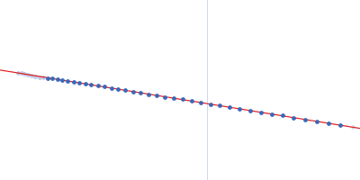
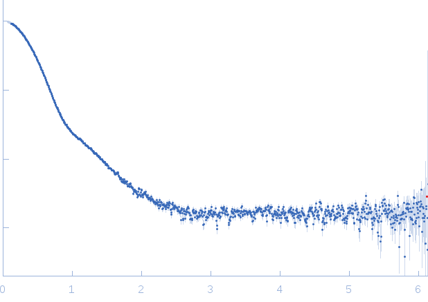
X-ray synchrotron radiation scattering data from solutions of the complex between GM-CSF/IL-2 inhibition factor and ovine Interleukin-2 in 20 mM HEPES 150 mM NaCl were collected on the SWING camera on the storage ring SOLEIL (Saint-Aubin, France) using a CCD AVIEX detector (I(s) vs s, where s = 4π sin θ/λ, where 2θ is the scattering angle). One solute concentration of  8.8 mg/ml was measured. 16 successive frames were collected. The data were normalized to the intensity of the transmitted beam and radially averaged; the scattering of the solvent-blank was subtracted and the different curves were scaled for protein concentration. The purity of the sample was obtained via SEC-SAXS.

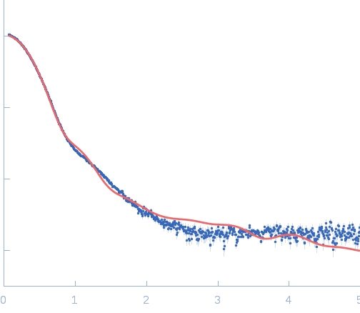
Modeling of SAXS data: A model of the GIF:IL-2 complex, based on the fit in a low-resolution (24 Å) map obtained by Negative-Stain Electron Microscopy, was used as an input for the Allosmod-FoXS web server to model missing loops, N- and C-termini and N-linked glycosylation. During each Allosmod-FoXS run, data up to a scattering angle of 0.5 Å-1 was used. Fits to the experimental SAXS data were calculated using the FoXS software. Note that the exact orientation of IL-2 is uncertain due to the low resolution of the EM map.

GM-CSF/IL-2 inhibition factor
Mol. type   Protein
Organism   Orf virus
Olig. state   Tetramer
Mon. MW   29.9 kDa
 
UniProt   Q9J5U5
Sequence   FASTA
 
Interleukin-2
Mol. type   Protein
Organism   Ovis aries
Olig. state   Monomer
Mon. MW   15.6 kDa
 
UniProt   P19114 (21-155)
Sequence   FASTA