|
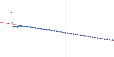
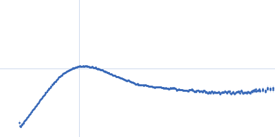
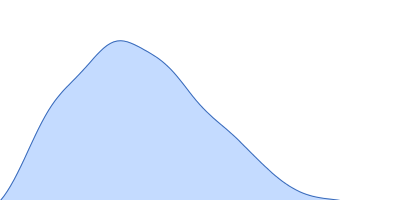
X-ray synchrotron radiation scattering data from solutions of Macrophage colony-stimulating factor 1 in complex with Macrophage colony-stimulating factor 1 receptor in 50 mM NaH2PO4, 100 mM NaCl (pH 7.4), were collected on the X33 camera on the storage ring DORIS (Hamburg, Germany). Using a 2D Photon counting Pilatus 1M-W pixel detector (s = 4π sin θ/λ, where 2θ is the scattering angle). Different solute concentrations in the range 0.50-10.00 mg/ml were measured. 15 successive 8 second frames were collected. The data were normalized to the intensity of the transmitted beam and radially averaged; the scattering of the solvent-blank was subtracted and the different curves were scaled for protein concentration. The low angle data collected were merged with the highest concentration high angle data to yield the final composite scattering curve.
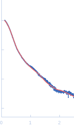
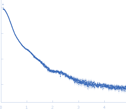
SAXS data acquisition for the glycosylated ternary hCSF-1:hCSF-1R full ectodomain complex (MW including glycans: 158 kDa) and subsequent data processing were performed as described in Elegheert et al., 2011 and Felix et al., 2013 respectively (see references). Rigid-body refinements of the hCSF-1:hCSF-1R complex were performed using the online version of SASREF. For each run, data to 0.25 angstrom-1 was used while imposing P2 symmetry. The crystal structure of full length hCSF-1:hCSF-1R without D1 was taken as a starting rigid-body core (PDB ID: 4WRM). D1 was added as a separate rigid body with a contact restraint of 4 angstrom between its C-terminus and the N-terminus of hCSF-1RD2-D5, as well as 5 N-linked glycans containing a 'dummy' Asparagine residue (Asn-GlcNac2Man5) with 1 angstrom restraints to the C-alpha of truncated Asparagine residues on hCSF-1RD2-D5 (N73, N153, N240, N275 and N353). The SASREF calculated fit of the model to the experimental data gave a chi-squared of 1.65, recalculation of the fit using Crysol and FoXS gave a chi-squared of 3.0 and 1.5 respectively. Here, the fit calculated with FoXS is presented. Felix, J., Elegheert, J., Gutsche, I., Shkumatov, A.V., Wen, Y., Bracke, N., Pannecoucke, E., Vandenberghe, I., Devreese, B., Svergun, D.I., et al. (2013). Human IL-34 and CSF-1 establish structurally similar extracellular assemblies with their common hematopoietic receptor. Structure 21, 528-539. Elegheert, J., Desfosses, A., Shkumatov, A.V., Wu, X., Bracke, N., Verstraete, K., Van Craenenbroeck, K., Brooks, B.R., Svergun, D.I., Vergauwen, B., et al. (2011). Extracellular Complexes of the Hematopoietic Human and Mouse CSF-1 Receptor Are Driven by Common Assembly Principles. Structure 19, 1762-1772.
|
|
 s, nm-1