Human βB2-Crystallin Forms a Face-en-Face Dimer in Solution: An Integrated NMR and SAXS Study.

Xi Z, Whitley MJ, Gronenborn AM, Structure 25(3):496-505 (2017) Europe PMC

SASDBU9 – Terminally truncated human βB2-crystallin

Beta-crystallin B2
MWexperimental 39 kDa
MWexpected 42 kDa
VPorod 62 nm3
log I(s) 5.10×10⁻² 5.10×10⁻³ 5.10×10⁻⁴ 5.10×10⁻⁵
Beta-crystallin B2 small angle scattering data  s, nm-1
ln I(s)
Beta-crystallin B2 Guinier plot ln 5.10×10⁻² Rg: 2.1 nm 0 (2.1 nm)-2 s2
(sRg)2I(s)/I(0)
Beta-crystallin B2 Kratky plot 1.104 0 3 sRg
p(r)
Beta-crystallin B2 pair distance distribution function Rg: 2.1 nm 0 Dmax: 6.7 nm

Data validation


Fits and models


log I(s)
 s, nm-1
Beta-crystallin B2 DAMMIN model

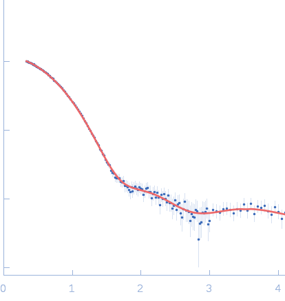
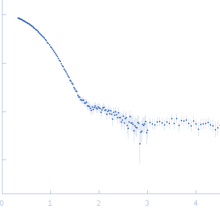
Synchrotron SAXS data from solutions of truncated human βB2-crystallin in 25 mM sodium phosphate, 5 mM DTT, 1 mM EDTA, pH 6.5, were collected on the 12ID-B SAXS/WAXS beam line at the Advanced Photon Source (APS, Argonne, IL, USA) using a Pilatus 2M detector at a sample-detector distance of 2 m and at a wavelength of λ = 0.08856 nm (I(s) vs s, where s = 4πsinθ/λ and 2θ is the scattering angle). Data were acquired over 30 successive 0.400 second frames from a sample at 1.75 mg/ml. The data were normalized to the intensity of the transmitted beam and radially averaged. The scattering of the radially-averaged solvent-blank was subtracted to produce the scattering profile displayed in this entry.

Cell temperature = UNKNOWN. Storage temperature = UNKNOWN

Beta-crystallin B2
Mol. type   Protein
Organism   Homo sapiens
Olig. state   Dimer
Mon. MW   20.9 kDa
 
UniProt   P43320 (15-195)
Sequence   FASTA