Synchrotron SAXS data from solutions of Dimeric Sortilin in the presence of neurotensin in 25 mM HEPES, 150 mM NaCl, pH 7.4, were collected at the BM29 beam line at the ESRF storage ring (Grenoble, France) using a Pilatus 1M detector at a sample-detector distance of 2.9 m and at a wavelength of λ = 0.099 nm (I(s) vs s, where s = 4πsinθ/λ and 2θ is the scattering angle). The data were collected at at 20°C. The data were normalized to the intensity of the transmitted beam and radially averaged; the scattering of the solvent-blank was subtracted.
Number of frames used for averaging: unknown. Exposure time: unknown. Sample concentration: unknown.
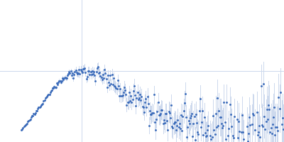
 s, nm-1
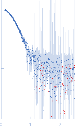