Solution structure of the Atg1 complex: implications for the architecture of the phagophore assembly site.

Köfinger J, Ragusa MJ, Lee IH, Hummer G, Hurley JH, Structure 23(5):809-818 (2015) Europe PMC

SASDCL3 – Atg1-Atg13 Subcomplex

Serine/threonine-protein kinase ATG1
Autophagy-related protein 13
MWexperimental 79 kDa
MWexpected 79 kDa
VPorod 160 nm3
log I(s) 1.01×10³ 1.01×10² 1.01×101 1.01×100
Serine/threonine-protein kinase ATG1 Autophagy-related protein 13 small angle scattering data  s, nm-1
ln I(s)
Serine/threonine-protein kinase ATG1 Autophagy-related protein 13 Guinier plot ln 1.02×10³ Rg: 3.3 nm 0 (3.3 nm)-2 s2
(sRg)2I(s)/I(0)
Serine/threonine-protein kinase ATG1 Autophagy-related protein 13 Kratky plot 1.104 0 3 sRg
p(r)
Serine/threonine-protein kinase ATG1 Autophagy-related protein 13 pair distance distribution function Rg: 3.3 nm 0 Dmax: 10.9 nm

Data validation


There are no models related to this curve.

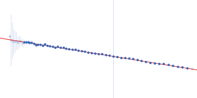
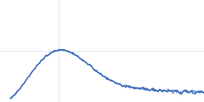
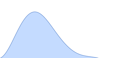
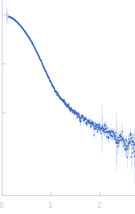
Synchrotron SAXS data from solutions of the Atg1-Atg13 Subcomplex in 20 mM Tris, 200 mM NaCl, 2% glycerol, pH 8 were collected on the 12.3.1 (SIBYLS) beam line at the Advanced Light Source (ALS; Berkeley, CA, USA) using a Pilatus3 X 2M detector (I(s) vs s, where s = 4πsin θ/λ and 2θ is the scattering angle). Solute concentrations ranging between 1 and 5.2 mg/ml were measured at 22°C. The data were normalized to the intensity of the transmitted beam and radially averaged; the scattering of the solvent-blank was subtracted and the different curves were scaled for protein concentration.

Wavelength = UNKNOWN. Storage temperature = UNKNOWN. Sample detector distance = UNKNOWN. X-ray Exposure time = UNKNOWN. Number of frames = UNKNOWN

Serine/threonine-protein kinase ATG1 (Atg1)
Mol. type   Protein
Organism   Kluyveromyces lactis
Olig. state   Dimer
Mon. MW   30.7 kDa
 
UniProt   Q6CSX2 (562-831)
Sequence   FASTA
 
Autophagy-related protein 13 (Atg13)
Mol. type   Protein
Organism   Kluyveromyces lactis
Olig. state   Dimer
Mon. MW   8.7 kDa
 
UniProt   Q6CWK2 (400-475)
Sequence   FASTA