Identification of two p23 co-chaperone isoforms in Leishmania braziliensis exhibiting similar structures and Hsp90 interaction properties despite divergent stabilities.

Batista FA, Almeida GS, Seraphim TV, Silva KP, Murta SM, Barbosa LR, Borges JC, FEBS J 282(2):388-406 (2015) Europe PMC

SASDCV5 – Leishmania braziliensis p23A

Uncharacterized protein
MWexperimental 23 kDa
MWexpected 22 kDa
log I(s) 3.18×10⁻² 3.18×10⁻³ 3.18×10⁻⁴ 3.18×10⁻⁵
Uncharacterized protein small angle scattering data  s, nm-1
ln I(s)
Uncharacterized protein Guinier plot ln 3.18×10⁻² Rg: 3.3 nm 0 (3.3 nm)-2 s2
(sRg)2I(s)/I(0)
Uncharacterized protein Kratky plot 1.104 0 3 sRg
p(r)
Uncharacterized protein pair distance distribution function Rg: 3.4 nm 0 Dmax: 13 nm

Data validation


Fits and models


log I(s)
 s, nm-1
Uncharacterized protein DAMFILT model

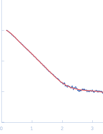
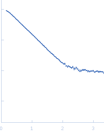
SAXS experiments were performed at the SAXS2 beamline in the Laboratório Nacional de Luz Síncrotron (LNLS, Campinas-SP, Brazil). The X-ray scattering data (I(s) vs s, where s = 4πsinθ/λ and 2θ is the scattering angle; λ = 0.1488 nm) were acquired using a two-dimensional MAR-CCD detector. Measurements were performed with a monochromatic X-ray beam and a sample-to-detector distance of ~1000 mm, corresponding to a scattering vector range of 0.015 < q < 0.35 Å−1. Scattering patterns were recorded at different sample concentrations (approximately 1.0, 2.0 and 4.0 mg/mL) and several frames in order to check for protein aggregation and X-ray damage. No evidence of aggregation or inter-particle interference were observed.

Number of frames = UNKNOWN

Uncharacterized protein (Lbp23A)
Mol. type   Protein
Organism   Leishmania braziliensis
Olig. state   Monomer
Mon. MW   22.1 kDa
 
UniProt   A4HNB5
Sequence   FASTA