Integrative dynamic structural biology unveils conformers essential for the oligomerization of a large GTPase.

Peulen TO, Hengstenberg CS, Biehl R, Dimura M, Lorenz C, Valeri A, Folz J, Hanke CA, Ince S, Vöpel T, Farago B, Gohlke H, Klare JP, Stadler AM, Seidel CAM, Herrmann C, Elife 12 (2023) Europe PMC

SASDDD6 – Human Guanylate-binding protein (hGBP1)

Guanylate-binding protein 1
MWexperimental 65 kDa
MWexpected 68 kDa
VPorod 105 nm3
log I(s) 6.59×101 6.59×100 6.59×10⁻¹ 6.59×10⁻²
Guanylate-binding protein 1 small angle scattering data  s, nm-1
ln I(s)
Guanylate-binding protein 1 Guinier plot ln 6.59×101 Rg: 3.9 nm 0 (3.9 nm)-2 s2
(sRg)2I(s)/I(0)
Guanylate-binding protein 1 Kratky plot 1.104 0 3 sRg
p(r)
Guanylate-binding protein 1 pair distance distribution function Rg: 4.0 nm 0 Dmax: 14.4 nm

Data validation


Fits and models


log I(s)
 s, nm-1
Guanylate-binding protein 1 DAMMIF model

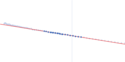
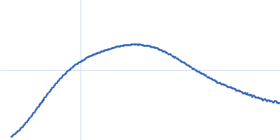
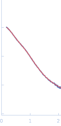
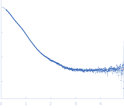
Synchrotron SAXS data from solutions of Human Guanylate-binding protein (hGBP1) in 50 mM TRIS, 5 mM MgCl2, 150 mM NaCl, pH 7.9 were collected using SEC-SAXS on the BM29 beam line at the ESRF (Grenoble, France) using a Pilatus 1M detector at a sample-detector distance of 2.9 m and at a wavelength of λ = 0.099 nm (l(s) vs s, where s = 4πsinθ/λ, and 2θ is the scattering angle). The SEC-column load concentration was 16.10 mg/ml (100 µl injection volume). 114 successive 1 second frames were collected through the elution peak, measured at 20°C. The data were normalized to the intensity of the transmitted beam and radially averaged; the scattering of an appropriate solvent-blank was subtracted.

Column type: Size exclusion chromatography (Superdex 200 10/300 GL, GE Healthcare) flow rate: 0.5 ml/min.

Guanylate-binding protein 1
Mol. type   Protein
Organism   Homo sapiens
Olig. state   Monomer
Mon. MW   67.6 kDa
 
UniProt   P32455
Sequence   FASTA