|
|
|
|
|
| Sample: |
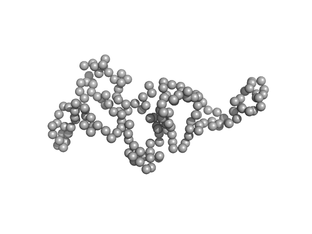
Disrupted- in-schizophrenia 1 (DISC1 12D2) 691-836 monomer, 19 kDa protein
|
| Buffer: |
25 mM Tris-HCl, 150 mM NaCl, 1mM DTT, pH: 7.4
|
| Experiment: |
SAXS
data collected at EMBL P12, PETRA III on 2015 Oct 1
|
Biophysical insights from a single chain camelid antibody directed against the Disrupted-in-Schizophrenia 1 protein.
PLoS One 13(1):e0191162 (2018)
...Stadler A, Köber S, Indurkhya X, Marreiros R, Trossbach SV, Bradshaw NJ, Prikulis I, Willbold D, Weiergräber OH, Korth C
|
| RgGuinier |
2.6 |
nm |
| Dmax |
7.3 |
nm |
| VolumePorod |
41 |
nm3 |
|