Integrin α6β4 Recognition of a Linear Motif of Bullous Pemphigoid Antigen BP230 Controls Its Recruitment to Hemidesmosomes.

Manso JA, Gómez-Hernández M, Carabias A, Alonso-García N, García-Rubio I, Kreft M, Sonnenberg A, de Pereda JM, Structure 27(6):952-964.e6 (2019) Europe PMC

SASDDN8 – Integrin beta4, fragment of the cytoplasmic region that includes the final part of the connecting segment and the third and fourth FnIII domains (FnIII-3,4). Point mutant R1542A

Integrin beta-4 (1436-1666) R1542A
MWexperimental 21 kDa
MWexpected 26 kDa
VPorod 34 nm3
log I(s) 1.52×10⁻² 1.52×10⁻³ 1.52×10⁻⁴ 1.52×10⁻⁵
Integrin beta-4 (1436-1666) R1542A small angle scattering data  s, nm-1
ln I(s)
Integrin beta-4 (1436-1666) R1542A Guinier plot ln 1.52×10⁻² Rg: 2.3 nm 0 (2.3 nm)-2 s2
(sRg)2I(s)/I(0)
Integrin beta-4 (1436-1666) R1542A Kratky plot 1.104 0 3 sRg
p(r)
Integrin beta-4 (1436-1666) R1542A pair distance distribution function Rg: 2.3 nm 0 Dmax: 8.5 nm

Data validation


Fits and models


log I(s)
 s, nm-1
Integrin beta4, fragment of the cytoplasmic region that includes the final part of the connecting segment and the third and fourth FnIII domains (FnIII-3,4). Point mutant R1542A Rg histogram Rg, nm

log I(s)
 s, nm-1
Integrin beta-4 (1436-1666) R1542A DAMMIF model

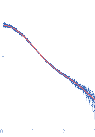
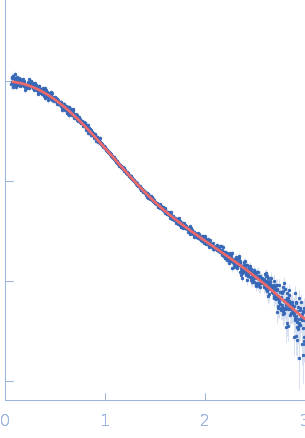
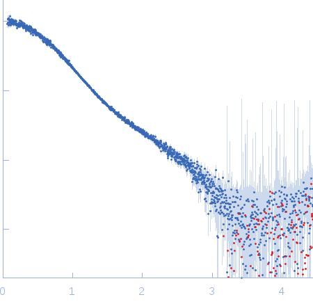
Synchrotron SAXS data from solutions of the integrin beta4 fragment (point mutant R1542A) in 20 mM sodium phosphate 150 mM NaCl 5% glycerol 3 mM DTT, pH 7.5 were collected on the EMBL P12 beam line at the PETRA III storage ring (Hamburg, Germany) using a Pilatus 2M detector at a sample-detector distance of 3.1 m and at a wavelength of λ = 0.124 nm (l(s) vs s, where s = 4πsinθ/λ, and 2θ is the scattering angle). Solute concentrations ranging between 0.9 and 14.5 mg/ml were measured at 10°C. 30 successive 0.050 second frames were collected. The data were normalized to the intensity of the transmitted beam and radially averaged; the scattering of the solvent-blank was subtracted. The low angle data collected at lower concentration were merged with the highest concentration high angle data to yield the final composite scattering curve.

Synchrotron SAXS data from solutions of a fragment of the cytoplasmic region of the human integrin Beta4 subunit. This fragment includes the final part of a region named the "connecting segment" the third and fourth fibronectin type III domains (FnIII-3,4). The protein contains the point mutation R1542A. Samples at different solute concentration in the range 0.9 – 14.5 mg/ml were obtained by 2-fold serial dilution and were measured.

Integrin beta-4 (1436-1666) R1542A (B4 CS-FnIII-3,4)
Mol. type   Protein
Organism   Homo sapiens
Olig. state   Monomer
Mon. MW   25.5 kDa
 
UniProt   P16144-2 (1436-1666)
Sequence   FASTA