|
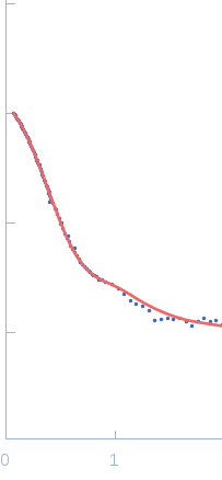
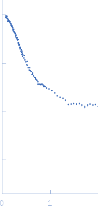
SANS data from solutions of Glutamate receptor 2 (GluA2) in the AMPA bound state, at neutral pH in D2O based buffer. 1 mM AMPA, 20 mM Tris/DCl, 100 mM NaCl, 0.5 mM deuterated n-dodecyl-β-D-maltopyranoside (synthesized to match out at 100% D2O), pH 7.5 were collected on the KWS1 camera at the FRM2 storage ring (Munich, Germany) using a 6Li-Scintillator 1 mm thickness + photomultiplier detector at a wavelength of λ = 0.5 nm (l(s) vs s, where s = 4πsinθ/λ, and 2θ is the scattering angle). One solute concentration of 0.31 mg/ml was measured at 10°C. 15 successive 900 second frames were collected. The data were normalized to the intensity of the transmitted beam and radially averaged; the scattering of the solvent-blank was subtracted.
Sample of GluA2 in the AMPA bound state at neutral pH. The SANS data were fitted with a mixture of GluA2 in a tetrameric compact form and a fraction of oligimers of the compact tetramer. Three models fitted equally well (within a 5% significance level): a mixture of GluA2 in the activated state (pdb-code 5weo) and a fraction of oligomers (top fit: fit62.dat), a mixture of GluA2 in the desensitized state (pdb-code 5vhz) and a fraction of oligomers (middle fit: fit63.dat) and a mixture of GluA2 in the resting state (pdb-code 4u2p) and a fraction of oligomers (bottom fit: fit72.dat). The oligomers were described with a fractal structure factor (Teixeira, J. (1988). J. Appl. Crystallogr. 21, 781-785). Data were fitted with WillItFit (Pedersen, M. C., Arleth, L. & Mortensen, K. (2013). J. Appl. Crystallogr. 46, 1894-1898). The SANS data were measured in three settings (sample/collimation): 1.5m/4.m, 4m/4m, and 8m/8m. The attached data are merged into one data set with all three settings. Pair distance distribution functions were calculated with BayesApp (www.bayesapp.org). Due to relatively low protein concentration, the concentration measurement was inaccurate, and the MW was therefore evaluated by (concentration independent) Porod analysis using the Porod volume calculator implemented in PRIMUS, and with a volume-to-mass conversion constant of 0.83 kDa/nm^3 (Gekko, K. & Noguchi, H. (1979). J. Phys. Chem. 83, 2706-2714; Squire, P. G. & Himmel, M. E. (1979). Arch. Biochem. Biophys. 196, 165-177).
|
|
 s, nm-1
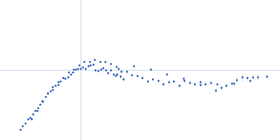