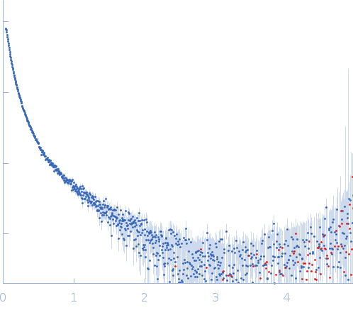
Synchrotron SAXS data from solutions of the polymeric form of Wag31 T73E in 50mM Tris, 300mM NaCl, 10% glycerol, 1mM EDTA (ethylene diamine tetra acetic acid), 5mM β-mercaptoethanol (BME), pH 7.5 were collected on the BM29 beam line at the ESRF (Grenoble, France) using a Pilatus 1M detector at a sample-detector distance of 2.9 m and at a wavelength of λ = 0.1 nm (l(s) vs s, where s = 4πsinθ/λ, and 2θ is the scattering angle). One solute concentration of 2.70 mg/ml was measured at 4°C. 10 successive 1 second frames were collected. The data were normalized to the intensity of the transmitted beam and radially averaged; the scattering of the solvent-blank was subtracted.
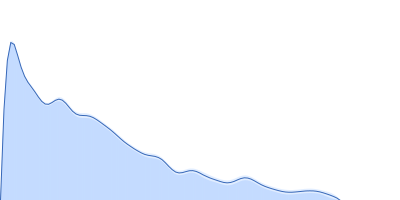
The cell wall synthesis protein Wag31: Polymer tbWag31 T73E forms a large super-macromolecular assembly (supported by atomic force microscopy, AFM) and as a result there is a very limited angular range to estimate the radius of gyration, Rg, using the Guinier approximation. In addition, the quoted monomer molecular weight does not apply to this sample. The probable distribution of real-space scattering pair vector lengths (p(r) vs r profile or PDDF) represents the cross-sectional pair distribution function for rod-like particle (option 4 in GNOM) and the subsequent data validation tools for this entry are based on the corresponding cross section parameters (Rg of cross section and Dmax of cross section). The I(s) is recorded in a relative scale in kDa units assuming a 1 mg/ml sample concentration. However the experimental concentration was found to be 2.7 mg/ml (assessed using a Bradford assay).
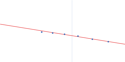
 s, nm-1
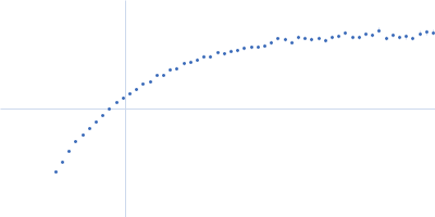