Regulation of futile ligation during early steps of BER in M. tuberculosis is carried out by a β-clamp-XthA-LigA tri-component complex

Shukla A, Afsar M, Khanam T, Kumar N, Ali F, Kumar S, Jahan F, Ramachandran R, International Journal of Biological Macromolecules (2022) DOI

SASDFH6 – Probable exodeoxyribonuclease III protein XthA, sliding beta clamp, DNA ligase-A tricomplex in the presence of DNA substrate

Probable exodeoxyribonuclease III protein XthA
DNA ligase A
Beta sliding clamp
DNA ligase A nicked DNA substrate
MWexperimental 214 kDa
MWexpected 209 kDa
VPorod 496 nm3
log I(s) 1.36×10² 1.36×101 1.36×100 1.36×10⁻¹
Probable exodeoxyribonuclease III protein XthA DNA ligase A Beta sliding clamp DNA ligase A nicked DNA substrate small angle scattering data  s, nm-1
ln I(s)
Probable exodeoxyribonuclease III protein XthA DNA ligase A Beta sliding clamp DNA ligase A nicked DNA substrate Guinier plot ln 1.37×10² Rg: 5.8 nm 0 (5.8 nm)-2 s2
(sRg)2I(s)/I(0)
Probable exodeoxyribonuclease III protein XthA DNA ligase A Beta sliding clamp DNA ligase A nicked DNA substrate Kratky plot 1.104 0 3 sRg
p(r)
Probable exodeoxyribonuclease III protein XthA DNA ligase A Beta sliding clamp DNA ligase A nicked DNA substrate pair distance distribution function Rg: 5.8 nm 0 Dmax: 19.1 nm

Data validation


Fits and models


log I(s)
 s, nm-1
Probable exodeoxyribonuclease III protein XthA DNA ligase A Beta sliding clamp DNA ligase A nicked DNA substrate DAMMIF model

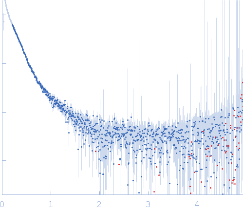
Synchrotron SAXS data from solutions of the XthA, sliding beta clamp, DNA ligase-A tricomplex with DNA substrate in 50 mM Tris pH 8.0, 200 mM NaCl , 2 mM β-mercaptoethanol, were collected on the BM29 beam line at the ESRF storage ring (Grenoble, France) using a Pilatus 1M detector at a sample-detector distance of 2.9 m and at a wavelength of λ = 0.0992 nm (I(s) vs s, where s = 4πsinθ/λ, and 2θ is the scattering angle). One solute concentration of 2.50 mg/ml was measured at 10°C. 10 successive 1 second frames were collected. The data were normalized to the intensity of the transmitted beam and radially averaged; the scattering of the solvent-blank was subtracted.

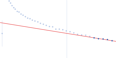
The presented model shows the spatial alignment of several individually calculated models and does not fit the SAXS data. A corresponding individual model fit (individual model not provided for this entry) is shown. The sample is severely aggregated.
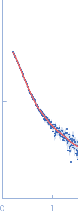
Probable exodeoxyribonuclease III protein XthA (MtbXthA)
Mol. type   Protein
Organism   Mycobacterium tuberculosis
Olig. state   Monomer
Mon. MW   30.7 kDa
 
UniProt   A0A0T9L251 (1-289)
Sequence   FASTA
 
DNA ligase A
Mol. type   Protein
Organism   Mycobacterium tuberculosis
Olig. state   Monomer
Mon. MW   76.1 kDa
 
UniProt   P9WNV1
Sequence   FASTA
 
Beta sliding clamp
Mol. type   Protein
Organism   Mycobacterium tuberculosis
Olig. state   Dimer
Mon. MW   42.9 kDa
 
UniProt   P9WNU1 (1-402)
Sequence   FASTA
 
DNA ligase A nicked DNA substrate
Mol. type   DNA
Olig. state   Dimer
Mon. MW   8.1 kDa
Sequence   FASTA