Dmax unknown – experimental data range validation not possible.
There are no models related to this curve.
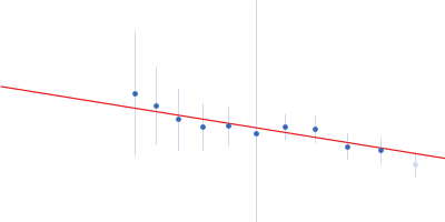
Synchrotron SAXS data from solutions of Cell wall synthesis protein Wag31(T73A) polymer (SEC-frames 300-304) in 50mM Tris pH7.5, 300mM NaCl, 10% Glycerol, 1mM EDTA (ethylene diamine tetra acetic acid), 5mM β-mercaptoethanol (BME), pH 7.5 were collected on the BL4-2 beam line at the Stanford Synchrotron Radiation Lightsource (SSRL) storage ring (Stanford, CA, USA) using a Rayonix MX225-HE detector at a sample-detector distance of 2.5 m and at a wavelength of λ = 0.1 nm (l(s) vs s, where s = 4πsinθ/λ, and 2θ is the scattering angle). One solute concentration of 2.40 mg/ml was measured at 23°C. 500 successive 1 second frames were collected. The data were normalized to the intensity of the transmitted beam and radially averaged; the scattering of the solvent-blank was subtracted.
SEC-SAXS was performed at room temperature using the following parameters: Column: Superdex 200 PC3.2; Flow rate: 0.05 mL/min; Sample injection concentration: 2.4 mg/mL; Injection volume: 100 μL. 500 successive 1 second frames were collected through the SEC-elution peak.
SEC-SAXS data of Cell wall synthesis protein Wag31(T73A) polymer frame no. 300-304
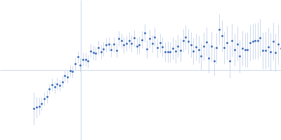
 s, nm-1
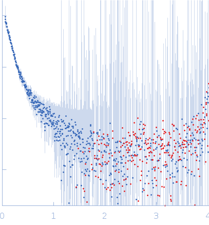